

Aim:
The present study aims to analyze the impact on cognitive recovery of an interdisciplinary treatment for acute and severe psychiatric patients.
Methods:
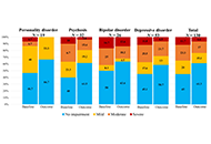
The present research is a naturalistic observational study of 130 adults (mean age of 47.68 years, 68% women). Clinical severity was assessed using Brief Psychiatry Rating Scale (BPRS), Montgomery-Asberg Depression Rating Scale (MADRS), and Hamilton Anxiety Rating Scale (HARS). Functional performance was evaluated using the Functioning Assessment Short Test (FAST), and cognitive impairment by applying the Montreal Cognitive Assessment (MoCA). Patients were clustered into four diagnostic groups (non-affective psychosis, bipolar, depressive, and personality disorders) and had individualized psychopharmacological treatment. They receive a transdiagnostic group program including several interventions that have shown evidence of beneficial effects over the different cognitive domains impaired in mental illness (attention, speed of processing, memory, working memory, reasoning, and problem-solving), as well as social cognition domains (emotion processing and social skills), in combination with psychoeducation and some strategies oriented to achieve healthy lifestyle routines (balanced diet, physical exercise, sleep hygiene, and smoking and alcohol cessation).
Results:
All clinical scales scores were improved after the end of treatment compared with those achieved at admission (BPRS, MADRS, and HARS scores below the cut-off point for establishing a case diagnosis). MoCA scores improved after the end of treatment concerning admission, both in the total score and in the differentiated cognitive domains, excluding orientation, which remained unchanged in the whole of the sample studied. No statistical significance was found in any comparisons between different diagnostic groups. No correlation between MoCA scores and BPRS, MADRS, or HARS scores at admission or discharge was found.
Conclusions:
These results show that the interdisciplinary therapeutic intervention can be effective for recovering cognitive impairment associated with mental disorders, irrespective of the diagnosis.
Aim:
The present study aims to analyze the impact on cognitive recovery of an interdisciplinary treatment for acute and severe psychiatric patients.
Methods:
The present research is a naturalistic observational study of 130 adults (mean age of 47.68 years, 68% women). Clinical severity was assessed using Brief Psychiatry Rating Scale (BPRS), Montgomery-Asberg Depression Rating Scale (MADRS), and Hamilton Anxiety Rating Scale (HARS). Functional performance was evaluated using the Functioning Assessment Short Test (FAST), and cognitive impairment by applying the Montreal Cognitive Assessment (MoCA). Patients were clustered into four diagnostic groups (non-affective psychosis, bipolar, depressive, and personality disorders) and had individualized psychopharmacological treatment. They receive a transdiagnostic group program including several interventions that have shown evidence of beneficial effects over the different cognitive domains impaired in mental illness (attention, speed of processing, memory, working memory, reasoning, and problem-solving), as well as social cognition domains (emotion processing and social skills), in combination with psychoeducation and some strategies oriented to achieve healthy lifestyle routines (balanced diet, physical exercise, sleep hygiene, and smoking and alcohol cessation).
Results:
All clinical scales scores were improved after the end of treatment compared with those achieved at admission (BPRS, MADRS, and HARS scores below the cut-off point for establishing a case diagnosis). MoCA scores improved after the end of treatment concerning admission, both in the total score and in the differentiated cognitive domains, excluding orientation, which remained unchanged in the whole of the sample studied. No statistical significance was found in any comparisons between different diagnostic groups. No correlation between MoCA scores and BPRS, MADRS, or HARS scores at admission or discharge was found.
Conclusions:
These results show that the interdisciplinary therapeutic intervention can be effective for recovering cognitive impairment associated with mental disorders, irrespective of the diagnosis.
DOI: https://doi.org/10.37349/ent.2023.00051
Aim:
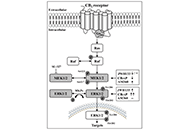
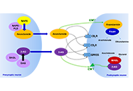
The sequential phosphorylation of mitogen-activated protein (MAP) kinases MEK-ERK is the most relevant cellular signaling pathway. This study quantified the parallel in vivo regulation of brain phosphorylation-MEK1/2 (p-MEK1/2) to p-ERK1/2 by mechanistically different cannabinoid 2 (CB2) receptor ligands, i.e., direct (and endogenous) agonists and inverse agonists.
Methods:
Groups of Swiss albino CD1 IGS male adult mice were treated (i.p.) with the CB2 agonist JWH133 (1 mg/kg and 3 mg/kg, 1 h, n = 8) or the CB2 inverse agonist/antagonist AM630 (0.3 mg/kg and 1 mg/kg, 1.5 h, n = 8–9), and 0.9% NaCl (2 mL/kg, 1 h, n = 4–10) as vehicle control. Transgenic male mice overexpressing cortical CB2 receptors [messenger RNA (mRNA) and protein] on a Swiss ICR congenic background (CB2xP) and the corresponding littermates age-matched wild-type (WT) controls were used. Protein forms (total MEK and ERK p-kinases) were resolved by electrophoresis [sodium dodecyl-sulfate polyacrylamide gel electrophoresis (SDS-PAGE) minigels] followed by immunoblotting standard procedures.
Results:
The selective CB2 agonist JWH133 (1 mg/kg and 3 mg/kg, i.p., 1 h) modestly decreased MEK (17%, n = 8) and upregulated ERK (25%, n = 8) activities. The endogenous CB2 agonists (acting on promoted overexpressed receptors) decreased MEK (44%, n = 9) and upregulated ERK (67%, n = 10) activities. The inverse agonist/antagonist AM630 (0.3 mg/kg and 1 mg/kg, i.p., 1.5 h) increases MEK activity (27%, n = 8) without significantly altering that of ERK (5%, n = 9).
Conclusions:
Acute treatments of mice with mechanistically different CB2 receptor ligands (i.e., direct agonists, endogenous agonists, and inverse agonists) resulted in disruption of MEK (p-MEK/total-MEK ratio) to ERK (p-ERK/total-ERK ratio) signals in the brain cortex. This striking disruption of MEK to ERK parallel regulation in the cannabinoid CB2 receptor system in the brain could be relevant to the postulated role of CB2 receptors in various central nervous system (CNS) diseases.
Aim:
The sequential phosphorylation of mitogen-activated protein (MAP) kinases MEK-ERK is the most relevant cellular signaling pathway. This study quantified the parallel in vivo regulation of brain phosphorylation-MEK1/2 (p-MEK1/2) to p-ERK1/2 by mechanistically different cannabinoid 2 (CB2) receptor ligands, i.e., direct (and endogenous) agonists and inverse agonists.
Methods:
Groups of Swiss albino CD1 IGS male adult mice were treated (i.p.) with the CB2 agonist JWH133 (1 mg/kg and 3 mg/kg, 1 h, n = 8) or the CB2 inverse agonist/antagonist AM630 (0.3 mg/kg and 1 mg/kg, 1.5 h, n = 8–9), and 0.9% NaCl (2 mL/kg, 1 h, n = 4–10) as vehicle control. Transgenic male mice overexpressing cortical CB2 receptors [messenger RNA (mRNA) and protein] on a Swiss ICR congenic background (CB2xP) and the corresponding littermates age-matched wild-type (WT) controls were used. Protein forms (total MEK and ERK p-kinases) were resolved by electrophoresis [sodium dodecyl-sulfate polyacrylamide gel electrophoresis (SDS-PAGE) minigels] followed by immunoblotting standard procedures.
Results:
The selective CB2 agonist JWH133 (1 mg/kg and 3 mg/kg, i.p., 1 h) modestly decreased MEK (17%, n = 8) and upregulated ERK (25%, n = 8) activities. The endogenous CB2 agonists (acting on promoted overexpressed receptors) decreased MEK (44%, n = 9) and upregulated ERK (67%, n = 10) activities. The inverse agonist/antagonist AM630 (0.3 mg/kg and 1 mg/kg, i.p., 1.5 h) increases MEK activity (27%, n = 8) without significantly altering that of ERK (5%, n = 9).
Conclusions:
Acute treatments of mice with mechanistically different CB2 receptor ligands (i.e., direct agonists, endogenous agonists, and inverse agonists) resulted in disruption of MEK (p-MEK/total-MEK ratio) to ERK (p-ERK/total-ERK ratio) signals in the brain cortex. This striking disruption of MEK to ERK parallel regulation in the cannabinoid CB2 receptor system in the brain could be relevant to the postulated role of CB2 receptors in various central nervous system (CNS) diseases.
DOI: https://doi.org/10.37349/ent.2023.00050
This article belongs to the special issue The Urgent Need for New Hypotheses to Develop Effective Therapeutic Tools Against Alzheimer's Disease
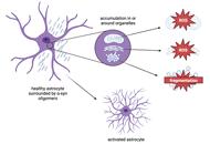
Post-translational modifications (PTMs) of alpha-synuclein (α-syn) can alter protein aggregation propensity to affect α-syn oligomer and fibril formation. The inflammatory response in Parkinson’s disease (PD) is mediated by microglia, astrocytes, T cells, B cells, macrophages, and neutrophils, which respond to α-syn aggregates in an attempt to clear synucleinopathy and restore brain homeostasis. This review focuses on the effects of PTMs on α-syn aggregation and cell-specific immune responses to α-syn aggregates in the context of PD.
Post-translational modifications (PTMs) of alpha-synuclein (α-syn) can alter protein aggregation propensity to affect α-syn oligomer and fibril formation. The inflammatory response in Parkinson’s disease (PD) is mediated by microglia, astrocytes, T cells, B cells, macrophages, and neutrophils, which respond to α-syn aggregates in an attempt to clear synucleinopathy and restore brain homeostasis. This review focuses on the effects of PTMs on α-syn aggregation and cell-specific immune responses to α-syn aggregates in the context of PD.
DOI: https://doi.org/10.37349/ent.2023.00052
Aim:
Parkinson’s disease (PD) is characterized by degeneration of midbrain dopamine neurons and synucleinopathy [aggregated alpha-synuclein protein (αSyn)]. The correlation between αSyn pathology and dopamine neuron degeneration remains to be fully established. Mouse models of PD are commonly used to increase knowledge of disease mechanisms. Lately, midbrain dopamine neurons have gained attention as more heterogeneous than previously recognized. With the aim to determine how the midbrain dopamine system in mice is affected in the presence of αSyn pathology, this brain system was studied in two transgenic mouse models of synucleinopathy.
Methods:
Brain sections from two previously described transgenic mouse lines verified for αSyn pathology through expression of the human αSyn gene (SNCA) under control of the Thy-1 promoter [Thy1-h[A30P]αSyn and Thy1-h[wt]αSyn (L61)], were analyzed using fluorescent in situ hybridization (FISH) and compared with matching sections from wild-type control mice. Probes directed towards mouse and human αSyn mRNA, and a battery of probes towards mRNAs representative of dopamine cell identity and heterogeneity, were implemented.
Results:
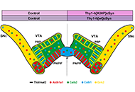
First, validation of αSyn-encoding mRNA was performed. Ample ectopic αSyn mRNA was observed throughout the brain of mice of each transgenic line. Next, midbrain dopamine neurons located in substantia nigra pars compacta (SNc) and ventral tegmental area (VTA) were analyzed using a battery of general and subpopulation-specific dopamine cell markers. This included tyrosine hydroxylase (Th), vesicular monoamine transporter 2 (Vmat2), dopamine transporter (Dat), aldehyde dehydrogenase 1 family member A1 (Aldh1a1), G-protein-activated inward-rectifying potassium channel type 2 (Girk2), calbindin 1 (Calb1), Calb2, gastrin-releasing peptide (Grp), and vesicular glutamate transporter 2 (Vglut2) mRNAs. No difference between transgenic and control mice was observed for any analyzed marker in either the Thy1-h[A30P]αSyn or Thy1-h[wt]αSyn transgenic mouse line.
Conclusions:
This study demonstrates remarkable robustness of midbrain dopamine cell integrity in the presence of brain-wide ectopic human αSyn in two transgenic mouse models of neurodegenerative disease, motivating further study into mechanisms correlating synucleinopathy with dopamine neuron degeneration in rodent models relevant to PD.
Aim:
Parkinson’s disease (PD) is characterized by degeneration of midbrain dopamine neurons and synucleinopathy [aggregated alpha-synuclein protein (αSyn)]. The correlation between αSyn pathology and dopamine neuron degeneration remains to be fully established. Mouse models of PD are commonly used to increase knowledge of disease mechanisms. Lately, midbrain dopamine neurons have gained attention as more heterogeneous than previously recognized. With the aim to determine how the midbrain dopamine system in mice is affected in the presence of αSyn pathology, this brain system was studied in two transgenic mouse models of synucleinopathy.
Methods:
Brain sections from two previously described transgenic mouse lines verified for αSyn pathology through expression of the human αSyn gene (SNCA) under control of the Thy-1 promoter [Thy1-h[A30P]αSyn and Thy1-h[wt]αSyn (L61)], were analyzed using fluorescent in situ hybridization (FISH) and compared with matching sections from wild-type control mice. Probes directed towards mouse and human αSyn mRNA, and a battery of probes towards mRNAs representative of dopamine cell identity and heterogeneity, were implemented.
Results:
First, validation of αSyn-encoding mRNA was performed. Ample ectopic αSyn mRNA was observed throughout the brain of mice of each transgenic line. Next, midbrain dopamine neurons located in substantia nigra pars compacta (SNc) and ventral tegmental area (VTA) were analyzed using a battery of general and subpopulation-specific dopamine cell markers. This included tyrosine hydroxylase (Th), vesicular monoamine transporter 2 (Vmat2), dopamine transporter (Dat), aldehyde dehydrogenase 1 family member A1 (Aldh1a1), G-protein-activated inward-rectifying potassium channel type 2 (Girk2), calbindin 1 (Calb1), Calb2, gastrin-releasing peptide (Grp), and vesicular glutamate transporter 2 (Vglut2) mRNAs. No difference between transgenic and control mice was observed for any analyzed marker in either the Thy1-h[A30P]αSyn or Thy1-h[wt]αSyn transgenic mouse line.
Conclusions:
This study demonstrates remarkable robustness of midbrain dopamine cell integrity in the presence of brain-wide ectopic human αSyn in two transgenic mouse models of neurodegenerative disease, motivating further study into mechanisms correlating synucleinopathy with dopamine neuron degeneration in rodent models relevant to PD.
DOI: https://doi.org/10.37349/ent.2023.00053
Astrocytes not only support neuronal function with essential roles in synaptic neurotransmission, action potential propagation, metabolic support, or neuroplastic and developmental adaptations. They also respond to damage or dysfunction in surrounding neurons and oligodendrocytes by releasing neurotrophic factors and other molecules that increase the survival of the supported cells or contribute to mechanisms of structural and molecular restoration. The neuroprotective responsiveness of astrocytes is based on their ability to sense signals of degeneration, metabolic jeopardy, and structural damage, and on their aptitude to locally deliver specific molecules to remedy threats to the molecular and structural features of their cellular partners. To the extent that neuronal and other glial cell disturbances are known to occur in affective disorders, astrocyte responsiveness to those disturbances may help to better understand the roles astrocytes play in affective disorders. The astrocytic sensing apparatus supporting those responses involves receptors for neurotransmitters, purines, cell adhesion molecules, and growth factors. Astrocytes also share with the immune system the capacity to respond to cytokines released upon neuronal damage. In addition, in response to specific signals, astrocytes release unique factors such as clusterin or humanin that have been shown to exert potent neuroprotective effects. Astrocytes integrate the signals above to further deliver structural lipids, remove toxic metabolites, stabilize the osmotic environment, normalize neurotransmitters, provide antioxidant protection, facilitate synaptogenesis, and act as barriers to contain varied deleterious signals, some of which have been described in brain regions relevant to affective disorders and related animal models. Since various injurious signals that activate astrocytes have been implicated in different aspects of the etiopathology of affective disorders, particularly in relation to the diagnosis of depression, potentiating the corresponding astrocyte neuroprotective responses may provide additional opportunities to improve or complement available pharmacological and behavioral therapies for affective disorders.
Astrocytes not only support neuronal function with essential roles in synaptic neurotransmission, action potential propagation, metabolic support, or neuroplastic and developmental adaptations. They also respond to damage or dysfunction in surrounding neurons and oligodendrocytes by releasing neurotrophic factors and other molecules that increase the survival of the supported cells or contribute to mechanisms of structural and molecular restoration. The neuroprotective responsiveness of astrocytes is based on their ability to sense signals of degeneration, metabolic jeopardy, and structural damage, and on their aptitude to locally deliver specific molecules to remedy threats to the molecular and structural features of their cellular partners. To the extent that neuronal and other glial cell disturbances are known to occur in affective disorders, astrocyte responsiveness to those disturbances may help to better understand the roles astrocytes play in affective disorders. The astrocytic sensing apparatus supporting those responses involves receptors for neurotransmitters, purines, cell adhesion molecules, and growth factors. Astrocytes also share with the immune system the capacity to respond to cytokines released upon neuronal damage. In addition, in response to specific signals, astrocytes release unique factors such as clusterin or humanin that have been shown to exert potent neuroprotective effects. Astrocytes integrate the signals above to further deliver structural lipids, remove toxic metabolites, stabilize the osmotic environment, normalize neurotransmitters, provide antioxidant protection, facilitate synaptogenesis, and act as barriers to contain varied deleterious signals, some of which have been described in brain regions relevant to affective disorders and related animal models. Since various injurious signals that activate astrocytes have been implicated in different aspects of the etiopathology of affective disorders, particularly in relation to the diagnosis of depression, potentiating the corresponding astrocyte neuroprotective responses may provide additional opportunities to improve or complement available pharmacological and behavioral therapies for affective disorders.
DOI: https://doi.org/10.37349/ent.2023.00054
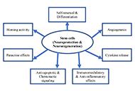
Self-neuronal regeneration is often limited or nonexistent after neuronal cell damage, making new technologies necessary for treating neurological damage. Although the brain can partially compensate by increasing its plasticity, these compensatory mechanisms can never fully restore the pre-damage state. Analysis of the literature regarding stem cell therapy in case of neurological disorders. Stem cells have shown promise for treating various neurological disorders and disabilities due to their regenerative capacity. Transplanting or administration of different types of stem cells has yielded promising results in animal models and early clinical trials. However, concerns remain regarding their implementation. The type of stem cell used, the optimal method and route of administration, the number of stem cells administered, preconditioning, and the injection schedule all need to be determined. Additionally, the long-term safety of stem cell treatment and the recipient’s age requires further investigation. Despite these concerns, stem cell therapy holds tremendous promise for treating neurological disorders, and continued research and well-designed studies will be crucial for unlocking its full potential.
Self-neuronal regeneration is often limited or nonexistent after neuronal cell damage, making new technologies necessary for treating neurological damage. Although the brain can partially compensate by increasing its plasticity, these compensatory mechanisms can never fully restore the pre-damage state. Analysis of the literature regarding stem cell therapy in case of neurological disorders. Stem cells have shown promise for treating various neurological disorders and disabilities due to their regenerative capacity. Transplanting or administration of different types of stem cells has yielded promising results in animal models and early clinical trials. However, concerns remain regarding their implementation. The type of stem cell used, the optimal method and route of administration, the number of stem cells administered, preconditioning, and the injection schedule all need to be determined. Additionally, the long-term safety of stem cell treatment and the recipient’s age requires further investigation. Despite these concerns, stem cell therapy holds tremendous promise for treating neurological disorders, and continued research and well-designed studies will be crucial for unlocking its full potential.
DOI: https://doi.org/10.37349/ent.2023.00055
This article belongs to the special issue Therapeutic Targets for Neuroprotection in Ischemic Stroke
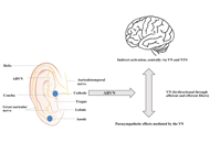
Vagus nerve stimulation (VNS) has gained prominence in the treatment of various clinical disorders such as migraine, depression, and tinnitus. Based on increased scientific knowledge of the VNS and insights into the vagus nerve (VN) function and anatomy/conduction, robust treatment approaches have been developed. There are both noninvasive and invasive VNS (iVNS) techniques. Currently, only iVNS techniques are approved by the US Food and Drug Administration (FDA). In contrast, transcutaneous VNS (tVNS) is a new treatment option that is receiving increasing attention. The tVNS application uses the cutaneous distribution of afferent VN fibers in the auricle, the auricular branch of the VN (ABVN), or in the neck, the cervical branch of the VN (CBVN). However, the tVNS technique has not yet been sufficiently researched in its application and mode of action to be used clinically on a large scale. Moreover, the stimulation parameters of the VN vary widely in different studies. Despite the growing number of research papers on this topic, more coherence in neurostimulation research and neuroanatomical basis is needed. The aim of this review is to highlight new clinical treatment options based on existing clinically applied treatment options. In this article, current clinical applications of tVNS are analyzed and important stimulation parameters are highlighted. Based on this data, useful new tVNS therapies are recommended. The focus will be placed on the study of inflammatory processes associated with cancer and on applications to cardiovascular events such as heart failure.
Vagus nerve stimulation (VNS) has gained prominence in the treatment of various clinical disorders such as migraine, depression, and tinnitus. Based on increased scientific knowledge of the VNS and insights into the vagus nerve (VN) function and anatomy/conduction, robust treatment approaches have been developed. There are both noninvasive and invasive VNS (iVNS) techniques. Currently, only iVNS techniques are approved by the US Food and Drug Administration (FDA). In contrast, transcutaneous VNS (tVNS) is a new treatment option that is receiving increasing attention. The tVNS application uses the cutaneous distribution of afferent VN fibers in the auricle, the auricular branch of the VN (ABVN), or in the neck, the cervical branch of the VN (CBVN). However, the tVNS technique has not yet been sufficiently researched in its application and mode of action to be used clinically on a large scale. Moreover, the stimulation parameters of the VN vary widely in different studies. Despite the growing number of research papers on this topic, more coherence in neurostimulation research and neuroanatomical basis is needed. The aim of this review is to highlight new clinical treatment options based on existing clinically applied treatment options. In this article, current clinical applications of tVNS are analyzed and important stimulation parameters are highlighted. Based on this data, useful new tVNS therapies are recommended. The focus will be placed on the study of inflammatory processes associated with cancer and on applications to cardiovascular events such as heart failure.
DOI: https://doi.org/10.37349/ent.2023.00056
This article belongs to the special issue Intervention of Neuroimmune Responses
Aim:
Stroke is the second most common cause of mortality and disability worldwide with ischemic strokes being the predominant type. The advent of neuroprotectants brought hope of improved outcomes and quality of life, but current guidelines, despite numerous trials, have no strong recommendation advising their use. This meta-analysis aims to evaluate the degree of effect and safety of the neuroprotectants cytidine-5’-diphosphocholine (CDP-choline), cerebrolysin, edaravone, and MLC601, in the recovery of patients with cerebral infarcts.
Methods:
An extensive literature search, through the databases of PubMed, PMC, Cochrane, and Ovid, was done with the keywords “CDP-choline”, “cerebrolysin”, “MLC601”, and “edaravone” each combined with the term “acute ischemic stroke”. Eligible studies included randomized controlled trials of these neuroprotectants administered to patients with acute ischemic strokes. A total of 2,025 studies were found, and after the application of screening criteria, 24 studies were eligible for analysis.
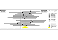
Results:
The analysis showed that the functional outcome of patients with acute ischemic strokes improved significantly when receiving neuroprotectants versus placebo supported by an odds ratio = 0.29 (0.09–0.50) with a confidence interval of 95%. The P-values are 0.0022 for the one-tailed test, and 0.0030 for the two-tailed test which express the significant improvement of functional outcomes in patients with acute ischemic strokes taking neuroprotectants.
Conclusions:
This study thus supports the use of neuroprotectants in patients with acute ischemic strokes to improve long-term functional outcomes and ultimately quality of life.
Aim:
Stroke is the second most common cause of mortality and disability worldwide with ischemic strokes being the predominant type. The advent of neuroprotectants brought hope of improved outcomes and quality of life, but current guidelines, despite numerous trials, have no strong recommendation advising their use. This meta-analysis aims to evaluate the degree of effect and safety of the neuroprotectants cytidine-5’-diphosphocholine (CDP-choline), cerebrolysin, edaravone, and MLC601, in the recovery of patients with cerebral infarcts.
Methods:
An extensive literature search, through the databases of PubMed, PMC, Cochrane, and Ovid, was done with the keywords “CDP-choline”, “cerebrolysin”, “MLC601”, and “edaravone” each combined with the term “acute ischemic stroke”. Eligible studies included randomized controlled trials of these neuroprotectants administered to patients with acute ischemic strokes. A total of 2,025 studies were found, and after the application of screening criteria, 24 studies were eligible for analysis.
Results:
The analysis showed that the functional outcome of patients with acute ischemic strokes improved significantly when receiving neuroprotectants versus placebo supported by an odds ratio = 0.29 (0.09–0.50) with a confidence interval of 95%. The P-values are 0.0022 for the one-tailed test, and 0.0030 for the two-tailed test which express the significant improvement of functional outcomes in patients with acute ischemic strokes taking neuroprotectants.
Conclusions:
This study thus supports the use of neuroprotectants in patients with acute ischemic strokes to improve long-term functional outcomes and ultimately quality of life.
DOI: https://doi.org/10.37349/ent.2023.00057
This article belongs to the special issue Therapeutic Targets for Neuroprotection in Ischemic Stroke
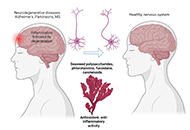
Recent investigations have shed light on the potential of seaweed, an abundant source of bioactive compounds, to mitigate and combat neurodegenerative diseases. In this comprehensive review, the accumulating evidence supporting the neuroprotective properties of seaweed-derived compounds is evaluated and their putative mechanisms of action are elucidated. The background of this review encompasses the general understanding of neurodegenerative diseases as debilitating conditions characterized by the progressive loss of nerve cell function and viability in the central nervous system. Furthermore, the global prevalence of these diseases, encompassing Alzheimer’s disease, Parkinson’s disease, and Huntington’s disease, and the persistent absence of effective treatments are emphasized. To address this critical issue, an innovative avenue of research is explored by investigating the potential of seaweed and its diverse array of bioactive compounds. By examining the available literature, the evidence supporting the neuroprotective effects of seaweed-derived compounds is consolidated. These bioactive constituents exhibit promising properties in preventing and mitigating neurodegeneration. Mechanistically, their actions involve intricate pathways that contribute to neuronal survival, reduction of oxidative stress, inhibition of neuroinflammation, and modulation of protein aggregation processes. This review provides a comprehensive analysis of the mechanisms underlying the neuroprotective effects of seaweed compounds. In conclusion, this review highlights the potential of seaweed as a valuable source of neuroprotective compounds and underscores the advancements made in this burgeoning field. The identification and elucidation of the mechanisms through which seaweed compounds exert their neuroprotective effects hold promise for the development of novel therapeutic interventions. These findings transcend disciplinary boundaries, offering insight into the potential application of seaweed-derived compounds as a valuable resource for combating neurodegenerative diseases across scientific domains.
Recent investigations have shed light on the potential of seaweed, an abundant source of bioactive compounds, to mitigate and combat neurodegenerative diseases. In this comprehensive review, the accumulating evidence supporting the neuroprotective properties of seaweed-derived compounds is evaluated and their putative mechanisms of action are elucidated. The background of this review encompasses the general understanding of neurodegenerative diseases as debilitating conditions characterized by the progressive loss of nerve cell function and viability in the central nervous system. Furthermore, the global prevalence of these diseases, encompassing Alzheimer’s disease, Parkinson’s disease, and Huntington’s disease, and the persistent absence of effective treatments are emphasized. To address this critical issue, an innovative avenue of research is explored by investigating the potential of seaweed and its diverse array of bioactive compounds. By examining the available literature, the evidence supporting the neuroprotective effects of seaweed-derived compounds is consolidated. These bioactive constituents exhibit promising properties in preventing and mitigating neurodegeneration. Mechanistically, their actions involve intricate pathways that contribute to neuronal survival, reduction of oxidative stress, inhibition of neuroinflammation, and modulation of protein aggregation processes. This review provides a comprehensive analysis of the mechanisms underlying the neuroprotective effects of seaweed compounds. In conclusion, this review highlights the potential of seaweed as a valuable source of neuroprotective compounds and underscores the advancements made in this burgeoning field. The identification and elucidation of the mechanisms through which seaweed compounds exert their neuroprotective effects hold promise for the development of novel therapeutic interventions. These findings transcend disciplinary boundaries, offering insight into the potential application of seaweed-derived compounds as a valuable resource for combating neurodegenerative diseases across scientific domains.
DOI: https://doi.org/10.37349/ent.2023.00058
This article belongs to the special issue Natural Products in Neurotherapeutic Applications
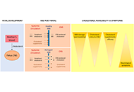
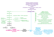
The autism spectrum disorder (ASD) comprises a series of neurological diseases that share serious alterations of the development of the central nervous system. The degree of disability may vary so that Asperger’s may have a relatively normal life and get positions of responsibility in corporations and even in Governments, whereas other ASD sufferers are fully dependent on caregivers and have serious cognitive deficits. Although the first cases of autism were detected by looking at failures in metabolism, e.g., phenylketonuria, to later identify the faulty gene, today the trend is the opposite, first obtaining the exome and minimizing the look for altered parameters in blood, urine, etc. Cholesterol is key for neural development as it is not able to cross the blood brain barrier. Therefore, any gene or environmental factor that affects cholesterol synthesis will impact early developmental stages eventually leading to a disease within the autism spectrum and/or schizophrenia. This review provides data of the relevance of cholesterol dyshomeostasis in autism spectrum disorders. Determining biochemical parameters in body fluids should help to provide new therapeutic approaches in some cases of autism.
The autism spectrum disorder (ASD) comprises a series of neurological diseases that share serious alterations of the development of the central nervous system. The degree of disability may vary so that Asperger’s may have a relatively normal life and get positions of responsibility in corporations and even in Governments, whereas other ASD sufferers are fully dependent on caregivers and have serious cognitive deficits. Although the first cases of autism were detected by looking at failures in metabolism, e.g., phenylketonuria, to later identify the faulty gene, today the trend is the opposite, first obtaining the exome and minimizing the look for altered parameters in blood, urine, etc. Cholesterol is key for neural development as it is not able to cross the blood brain barrier. Therefore, any gene or environmental factor that affects cholesterol synthesis will impact early developmental stages eventually leading to a disease within the autism spectrum and/or schizophrenia. This review provides data of the relevance of cholesterol dyshomeostasis in autism spectrum disorders. Determining biochemical parameters in body fluids should help to provide new therapeutic approaches in some cases of autism.
DOI: https://doi.org/10.37349/ent.2021.00003
This article belongs to the special issue Cholesterol Dyshomeostasis in Neurological Diseases
Early in the course of infection, human immunodeficiency virus (HIV) is able to enter the central nervous system where it stablishes a permanent reservoir. Current antiretroviral therapies do not efficiently cross the blood-brain barrier and therefore do not reach the HIV located in the central nervous system. Consequently, HIV infection can often be associated with neurocognitive impairment and HIV-associated dementia. The purpose of this review is to brief the reader into the world of neurological complications arising from HIV infection. Mechanisms by which HIV directly or indirectly impairs the central nervous system are discussed, as well as other factors influencing or contributing to the impairment, and the animal models currently used to perform research on the topic.
Early in the course of infection, human immunodeficiency virus (HIV) is able to enter the central nervous system where it stablishes a permanent reservoir. Current antiretroviral therapies do not efficiently cross the blood-brain barrier and therefore do not reach the HIV located in the central nervous system. Consequently, HIV infection can often be associated with neurocognitive impairment and HIV-associated dementia. The purpose of this review is to brief the reader into the world of neurological complications arising from HIV infection. Mechanisms by which HIV directly or indirectly impairs the central nervous system are discussed, as well as other factors influencing or contributing to the impairment, and the animal models currently used to perform research on the topic.
DOI: https://doi.org/10.37349/ent.2021.00004
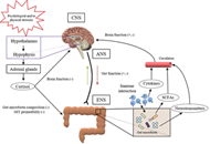
The pathogenic basis behind some of the most prevalent neurodegenerative diseases in advanced societies, known as proteinopathies, deals with alterations in protein homeostasis. Despite the broad diversity of clinical symptoms, they share a remarkably common feature that is the serious neuronal loss in several disease-specific brain regions due to the presence of toxic aggregations of misfolded proteins. So far, research efforts have been insufficient to decipher the exact molecular mechanisms that trigger the conformational change from a functional healthy protein to its pathological version. This is a sine qua non condition to progress in developing new approaches and treatments for these diseases for which there is no cure. Currently, it is well accepted that perturbations in gut microbiota composition negatively impact a wide range of brain processes via the gut-brain axis which increases host susceptibility to neurodegenerative disorders. In this context, modulate the microbial ecosystem colonizing the gastrointestinal tract may be a promising therapeutic approach in the management of proteinopathies. This review aims to provide an updated view of the role that gut microbiota poses in the pathogenesis of Parkinson’s disease, Alzheimer’s disease and Huntington’s disease, the most common neurodegenerative proteinopathies, and of the possibility of translating this knowledge into effective and safe clinical microbiota-based interventions, especially those designed to afford neuroprotection.
The pathogenic basis behind some of the most prevalent neurodegenerative diseases in advanced societies, known as proteinopathies, deals with alterations in protein homeostasis. Despite the broad diversity of clinical symptoms, they share a remarkably common feature that is the serious neuronal loss in several disease-specific brain regions due to the presence of toxic aggregations of misfolded proteins. So far, research efforts have been insufficient to decipher the exact molecular mechanisms that trigger the conformational change from a functional healthy protein to its pathological version. This is a sine qua non condition to progress in developing new approaches and treatments for these diseases for which there is no cure. Currently, it is well accepted that perturbations in gut microbiota composition negatively impact a wide range of brain processes via the gut-brain axis which increases host susceptibility to neurodegenerative disorders. In this context, modulate the microbial ecosystem colonizing the gastrointestinal tract may be a promising therapeutic approach in the management of proteinopathies. This review aims to provide an updated view of the role that gut microbiota poses in the pathogenesis of Parkinson’s disease, Alzheimer’s disease and Huntington’s disease, the most common neurodegenerative proteinopathies, and of the possibility of translating this knowledge into effective and safe clinical microbiota-based interventions, especially those designed to afford neuroprotection.
DOI: https://doi.org/10.37349/ent.2021.00005
Since the identification and cloning of the cannabinoid receptor 2 (CB2R), several studies focused on the characterization of its physiological and pathological role. Initially, CB2R was considered as the peripheral cannabinoid receptor due to its detection in the rat spleen and leukocyte subpopulation in humans. Later, CB2R was identified in different brain regions significantly modifying the landscape and pointing out its role in a wide variety of central physiological functions and pathological conditions. Additional research also detected the expression of CB2R in neurons, microglia, and astroglia in different brain regions. Indeed, the findings collected to date support a significant function of CB2R in anxiety, depression, schizophrenia, and additional neuropsychiatric disorders. This review gathers the most relevant literature regarding new advances about the role of CB2R in a variety of neuropsychiatric conditions, with special emphasis on its potential as a new therapeutic target for the treatment of different psychiatric disorders.
Since the identification and cloning of the cannabinoid receptor 2 (CB2R), several studies focused on the characterization of its physiological and pathological role. Initially, CB2R was considered as the peripheral cannabinoid receptor due to its detection in the rat spleen and leukocyte subpopulation in humans. Later, CB2R was identified in different brain regions significantly modifying the landscape and pointing out its role in a wide variety of central physiological functions and pathological conditions. Additional research also detected the expression of CB2R in neurons, microglia, and astroglia in different brain regions. Indeed, the findings collected to date support a significant function of CB2R in anxiety, depression, schizophrenia, and additional neuropsychiatric disorders. This review gathers the most relevant literature regarding new advances about the role of CB2R in a variety of neuropsychiatric conditions, with special emphasis on its potential as a new therapeutic target for the treatment of different psychiatric disorders.
DOI: https://doi.org/10.37349/ent.2021.00006
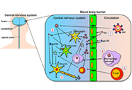
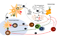
Current evidence indicates that neurodegeneration of dopaminergic neurons of the substantia nigra associated to Parkinson’s disease is a consequence of a neuroinflammatory process in which microglial cells play a central role. The initial activation of microglial cells is triggered by pathogenic protein inclusions, which are mainly composed by α-synuclein. Importantly, these pathogenic forms of α-synuclein subsequently induce a T-cell-mediated autoimmune response to dopaminergic neurons. Depending on their functional phenotype, these autoreactive T-cells might shape the functional features of activated microglia. T-cells bearing pro-inflammatory phenotypes such as T-helper (Th)1 or Th17 promote a chronic inflammatory behaviour on microglia, whilst anti-inflammatory T-cells, such as regulatory T-cells (Treg) favour the acquisition of neuroprotective features by microglia. Thus, T-cells play a fundamental role in the development of neuroinflammation and neurodegeneration involved in Parkinson’s disease. This review summarizes the evidence indicating that not only CD4+ T-cells, but also CD8+ T-cells play an important role in the physiopathology of Parkinson’s disease. Next, this review analyses the different T-cell epitopes derived from the pathogenic forms of α-synuclein involved in the autoimmune response associated to Parkinson’s disease in animal models and humans. It also summarizes the requirement of specific alleles of major histocompatibility complexes (MHC) class I and class II necessaries for the presentation of CD8+ and CD4+ T-cell epitopes from the pathogenic forms of α-synuclein in both humans and animal models. Finally, this work summarizes and discusses a number of experimental immunotherapies that aim to strengthen the Treg response or to dampen the inflammatory T-cell response as a therapeutic approach in animal models of Parkinson’s disease.
Current evidence indicates that neurodegeneration of dopaminergic neurons of the substantia nigra associated to Parkinson’s disease is a consequence of a neuroinflammatory process in which microglial cells play a central role. The initial activation of microglial cells is triggered by pathogenic protein inclusions, which are mainly composed by α-synuclein. Importantly, these pathogenic forms of α-synuclein subsequently induce a T-cell-mediated autoimmune response to dopaminergic neurons. Depending on their functional phenotype, these autoreactive T-cells might shape the functional features of activated microglia. T-cells bearing pro-inflammatory phenotypes such as T-helper (Th)1 or Th17 promote a chronic inflammatory behaviour on microglia, whilst anti-inflammatory T-cells, such as regulatory T-cells (Treg) favour the acquisition of neuroprotective features by microglia. Thus, T-cells play a fundamental role in the development of neuroinflammation and neurodegeneration involved in Parkinson’s disease. This review summarizes the evidence indicating that not only CD4+ T-cells, but also CD8+ T-cells play an important role in the physiopathology of Parkinson’s disease. Next, this review analyses the different T-cell epitopes derived from the pathogenic forms of α-synuclein involved in the autoimmune response associated to Parkinson’s disease in animal models and humans. It also summarizes the requirement of specific alleles of major histocompatibility complexes (MHC) class I and class II necessaries for the presentation of CD8+ and CD4+ T-cell epitopes from the pathogenic forms of α-synuclein in both humans and animal models. Finally, this work summarizes and discusses a number of experimental immunotherapies that aim to strengthen the Treg response or to dampen the inflammatory T-cell response as a therapeutic approach in animal models of Parkinson’s disease.
DOI: https://doi.org/10.37349/ent.2021.00007
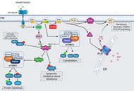
Phosphoinositides are membrane phospholipids involved in a variety of cellular processes like growth, development, metabolism, and transport. This review focuses on the maintenance of cellular homeostasis of phosphatidylinositol 4,5-bisphosphate (PIP2), and phosphatidylinositol 3,4,5-trisphosphate (PIP3). The critical balance of these PIPs is crucial for regulation of neuronal form and function. The activity of PIP2 and PIP3 can be regulated through kinases, phosphatases, phospholipases and cholesterol microdomains. PIP2 and PIP3 carry out their functions either indirectly through their effectors activating integral signaling pathways, or through direct regulation of membrane channels, transporters, and cytoskeletal proteins. Any perturbations to the balance between PIP2 and PIP3 signaling result in neurodevelopmental and neurodegenerative disorders. This review will discuss the upstream modulators and downstream effectors of the PIP2 and PIP3 signaling, in the context of neuronal health and disease.
Phosphoinositides are membrane phospholipids involved in a variety of cellular processes like growth, development, metabolism, and transport. This review focuses on the maintenance of cellular homeostasis of phosphatidylinositol 4,5-bisphosphate (PIP2), and phosphatidylinositol 3,4,5-trisphosphate (PIP3). The critical balance of these PIPs is crucial for regulation of neuronal form and function. The activity of PIP2 and PIP3 can be regulated through kinases, phosphatases, phospholipases and cholesterol microdomains. PIP2 and PIP3 carry out their functions either indirectly through their effectors activating integral signaling pathways, or through direct regulation of membrane channels, transporters, and cytoskeletal proteins. Any perturbations to the balance between PIP2 and PIP3 signaling result in neurodevelopmental and neurodegenerative disorders. This review will discuss the upstream modulators and downstream effectors of the PIP2 and PIP3 signaling, in the context of neuronal health and disease.
DOI: https://doi.org/10.37349/ent.2021.00008
This article belongs to the special issue Cholesterol Dyshomeostasis in Neurological Diseases
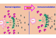
Spinal cord injury (SCI) induces several destructive events that develop immediately after the primary insult. These phenomena increase tissue damage; that is why, numerous therapeutic approaches are studied in order to neutralize these destructive mechanisms. In line with this, several studies indicate that after injury, neural tissue could be protected by an adaptive immune response directed against self-antigens. Immunization with neural-derived peptides (INDP) reduces secondary degeneration of neurons after spinal cord insult and promotes a significant motor recovery. The combination of antioxidants or other immunomodulatory peptides after SCI can improve the protective effect induced by INDP. INDP in acute SCI is a promising strategy, so further studies should be addressed to be able to formulate the best strategy.
Spinal cord injury (SCI) induces several destructive events that develop immediately after the primary insult. These phenomena increase tissue damage; that is why, numerous therapeutic approaches are studied in order to neutralize these destructive mechanisms. In line with this, several studies indicate that after injury, neural tissue could be protected by an adaptive immune response directed against self-antigens. Immunization with neural-derived peptides (INDP) reduces secondary degeneration of neurons after spinal cord insult and promotes a significant motor recovery. The combination of antioxidants or other immunomodulatory peptides after SCI can improve the protective effect induced by INDP. INDP in acute SCI is a promising strategy, so further studies should be addressed to be able to formulate the best strategy.
DOI: https://doi.org/10.37349/ent.2021.00009
Familial early-onset Alzheimer’s disease (AD) is more probable in individuals coming from mothers diagnosed with AD than from fathers diagnosed with AD. Studies in animal models have shown maternal imprinting due to the transmission to the embryo of altered material in the ovum. In the case of transgenic animals harboring a mutated form of the human amyloid precursor protein (APP), offspring from crosses with wild-type (WT) fathers and transgenic mothers display more abnormalities than offspring from crosses with transgenic fathers and WT mothers. Expression of the mutated APP in the ovum may lead to alterations that may be genetic and/or epigenetic in the nuclear and/or the mitochondrial DNA. These modifications that are transmitted to the new living beings affect more mitochondrial proteins and, therefore, the mitochondrial function may be affected in adulthood by trends present in the ovum.
Familial early-onset Alzheimer’s disease (AD) is more probable in individuals coming from mothers diagnosed with AD than from fathers diagnosed with AD. Studies in animal models have shown maternal imprinting due to the transmission to the embryo of altered material in the ovum. In the case of transgenic animals harboring a mutated form of the human amyloid precursor protein (APP), offspring from crosses with wild-type (WT) fathers and transgenic mothers display more abnormalities than offspring from crosses with transgenic fathers and WT mothers. Expression of the mutated APP in the ovum may lead to alterations that may be genetic and/or epigenetic in the nuclear and/or the mitochondrial DNA. These modifications that are transmitted to the new living beings affect more mitochondrial proteins and, therefore, the mitochondrial function may be affected in adulthood by trends present in the ovum.
DOI: https://doi.org/10.37349/ent.2021.00010
Peroxisomes are actively involved in the metabolism of various lipids including fatty acids, ether phospholipids, bile acids as well as the processing of reactive oxygen and nitrogen species. Recent studies show that peroxisomes can regulate cholesterol homeostasis by mediating cholesterol transport from the lysosomes to the endoplasmic reticulum and towards primary cilium as well. Disruptions of peroxisome biogenesis or functions lead to peroxisomal disorders that usually involve neurological deficits. Peroxisomal dysfunction is also linked to several neurodegenerative diseases such as Alzheimer’s disease and Parkinson’s disease. In many peroxisomal disorders and neurodegenerative diseases, aberrant cholesterol accumulation is frequently encountered yet largely neglected. This review discusses the current understanding of the mechanisms by which peroxisomes facilitate cholesterol trafficking within the cell and the pathological conditions related to impaired cholesterol transport by peroxisomes, with the hope to inspire future development of the treatments for peroxisomal disorders and neurodegenerative diseases.
Peroxisomes are actively involved in the metabolism of various lipids including fatty acids, ether phospholipids, bile acids as well as the processing of reactive oxygen and nitrogen species. Recent studies show that peroxisomes can regulate cholesterol homeostasis by mediating cholesterol transport from the lysosomes to the endoplasmic reticulum and towards primary cilium as well. Disruptions of peroxisome biogenesis or functions lead to peroxisomal disorders that usually involve neurological deficits. Peroxisomal dysfunction is also linked to several neurodegenerative diseases such as Alzheimer’s disease and Parkinson’s disease. In many peroxisomal disorders and neurodegenerative diseases, aberrant cholesterol accumulation is frequently encountered yet largely neglected. This review discusses the current understanding of the mechanisms by which peroxisomes facilitate cholesterol trafficking within the cell and the pathological conditions related to impaired cholesterol transport by peroxisomes, with the hope to inspire future development of the treatments for peroxisomal disorders and neurodegenerative diseases.
DOI: https://doi.org/10.37349/ent.2021.00011
This article belongs to the special issue Cholesterol Dyshomeostasis in Neurological Diseases
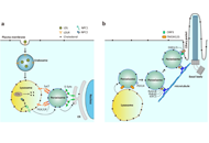
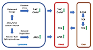
Niemann-Pick C disease is a rare neurodegenerative, lysosomal storage disease caused by accumulation of unesterified cholesterol. Diagnosis of the disease is often delayed due to its rarity, the heterogeneous presentation, and the early non-specific symptoms. The discovery of disease-specific biomarkers—cholestane-3β,5α,6β-triol (C-triol), trihydroxycholanic acid glycinate (TCG) and N-palmitoyl-O-phosphocholineserine [PPCS, initially referred to as lysosphingomyelin-509 (lysoSM-509)]—has led to development of non-invasive, blood-based diagnostics. Dissemination of these rapid, sensitive, and specific clinical assays has accelerated diagnosis. Moreover, the superior receiver operating characteristic of the TCG bile acid biomarker and its detection in dried blood spots has also facilitated development of a newborn screen for NPC, which is currently being piloted in New York state. The C-triol, TCG and PPCS biomarkers have also been proved useful for monitoring treatment response in peripheral tissues, but are uninformative with respect to treatment efficacy in the central nervous system (CNS). A major gap for the field is the lack of a validated, non-invasive biomarker to monitor the course of disease and CNS response to therapy.
Niemann-Pick C disease is a rare neurodegenerative, lysosomal storage disease caused by accumulation of unesterified cholesterol. Diagnosis of the disease is often delayed due to its rarity, the heterogeneous presentation, and the early non-specific symptoms. The discovery of disease-specific biomarkers—cholestane-3β,5α,6β-triol (C-triol), trihydroxycholanic acid glycinate (TCG) and N-palmitoyl-O-phosphocholineserine [PPCS, initially referred to as lysosphingomyelin-509 (lysoSM-509)]—has led to development of non-invasive, blood-based diagnostics. Dissemination of these rapid, sensitive, and specific clinical assays has accelerated diagnosis. Moreover, the superior receiver operating characteristic of the TCG bile acid biomarker and its detection in dried blood spots has also facilitated development of a newborn screen for NPC, which is currently being piloted in New York state. The C-triol, TCG and PPCS biomarkers have also been proved useful for monitoring treatment response in peripheral tissues, but are uninformative with respect to treatment efficacy in the central nervous system (CNS). A major gap for the field is the lack of a validated, non-invasive biomarker to monitor the course of disease and CNS response to therapy.
DOI: https://doi.org/10.37349/ent.2021.00012
This article belongs to the special issue Cholesterol Dyshomeostasis in Neurological Diseases
The brain cholesterol content is determined by the balance between the pathways of in situ biosynthesis and cholesterol elimination via 24-hydroxylation catalyzed by cytochrome P450 46A1 (CYP46A1). Both pathways are tightly coupled and determine the rate of brain cholesterol turnover. Evidence is accumulating that modulation of CYP46A1 activity by gene therapy or pharmacologic means could be beneficial in the case of neurodegenerative and other brain diseases and affect brain processes other than cholesterol biosynthesis and elimination. This minireview summarizes these other processes, most common of which include abnormal protein accumulation, memory, and cognition, motor behavior, gene transcription, protein phosphorylation as well as autophagy and lysosomal processing. The unifying mechanisms, by which these processes could be affected by CYP46A targeting are also discussed.
The brain cholesterol content is determined by the balance between the pathways of in situ biosynthesis and cholesterol elimination via 24-hydroxylation catalyzed by cytochrome P450 46A1 (CYP46A1). Both pathways are tightly coupled and determine the rate of brain cholesterol turnover. Evidence is accumulating that modulation of CYP46A1 activity by gene therapy or pharmacologic means could be beneficial in the case of neurodegenerative and other brain diseases and affect brain processes other than cholesterol biosynthesis and elimination. This minireview summarizes these other processes, most common of which include abnormal protein accumulation, memory, and cognition, motor behavior, gene transcription, protein phosphorylation as well as autophagy and lysosomal processing. The unifying mechanisms, by which these processes could be affected by CYP46A targeting are also discussed.
DOI: https://doi.org/10.37349/ent.2021.00013
This article belongs to the special issue Cholesterol Dyshomeostasis in Neurological Diseases
