


Editor's Picks
Open Access
Review
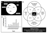
Advances in bone tissue engineering using biomaterial based scaffolds, purine crosslinking and Wnt signaling
The design of effective treatments for critical size bone defects, which result from various conditions such as trauma, infection, injury, or tumor resection, presents a significant challenge in clinical practice. While autologous grafts are commonly regarded as gold standard treatments in these complex healing scenarios, they are often associated with notable limitations, including donor site morbidity and limited graft volume. As a result, recent research trends have shifted towards developing biomaterials that better emulate the inherent complexity of the native bone structure and function through implementation of a “Diamond Concept” polytherapy strategy. Central to this approach is the utilization of biomaterials, increasingly composed of composite materials that integrate bioactive osteoinductive factors and cell sources to enhance healing outcomes. The usage of Wnt signaling specific agonists as osteoinductive mediators has been recently shown to be a promising strategy for promoting healing, as this pathway is well established to have an important role in both osteogenic differentiation and bone formation processes. Implementation of a localized delivery system through scaffold incorporation is necessary in this scenario, however, to minimize any potential off-target effects caused by the Wnt signaling cascade’s non-specificity to bone. Findings in the literature clearly show that this approach holds promise to improve clinical healing outcomes, paving the way for more effective treatment options. In this review, we will generally discuss the design of biomaterials, specifically bulk materials and composites, for the treatment of critical size bone defects. Additionally, we will highlight recent work on the design of chitosan-based scaffolds modified with purine crosslinking, to overcome cytotoxicity issues associated with other chemical crosslinkers. In this context, we focus on optimizing material design for this bone healing application and discuss the benefits of localized Wnt agonist as mediators to improve the scaffold’s osteoinductive behavior.
Open Access
Review
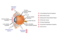
Advancements in hydrogel technology for ocular drug delivery
Hydrogel-based drug delivery systems (DDS) offer promising alternatives for treating ocular diseases by overcoming the limitations of traditional therapies, such as low bioavailability, frequent administration, and invasiveness. Hydrogels, with their high biocompatibility and ability to respond to external stimuli, can provide sustained and targeted drug delivery. This review highlights the unique properties of hydrogels, including their swelling behavior, porosity, and mechanical strength, making them suitable for various ocular applications. The classification of hydrogels based on cross-linking methods, origins, and stimuli responsiveness is discussed, emphasizing their potential in drug delivery for dry eye disease (DED), glaucoma, corneal alkali burns, and neovascularization. Notable advances include thermosensitive and pH-responsive hydrogels, which have shown promising results in preclinical studies. Despite these advances, most studies are still in preclinical stages, highlighting the need for rigorous human trials to validate the safety and efficacy of hydrogel DDS. Collaborative efforts among researchers, pharmacologists, and ophthalmologists are essential to translating these innovations into clinical practice, ultimately improving patient outcomes in ocular disease management.
Open Access
Commentary
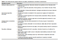
Navigating regulatory challenges in molecularly tailored nanomedicine
Nanomedicine, a convergence of nanotechnology and medical sciences, has unleashed transformative potential in healthcare. However, harnessing the benefits of nanomedicine requires a thorough understanding of its regulatory landscape. An in-depth discussion of regulatory considerations, including molecular safety assessment, harmonization of the regulatory landscape, and shaping the future of innovation, is presented in this discourse. The molecular safety assessment entails evaluating interactions between nanoparticles and biomolecules, ensuring compatibility at the molecular level. Harmonization involves developing international standards and guidelines for a consistent regulatory approach, while shaping innovations emphasizes integrating molecular safety assessments into early stages of development. Challenges encompass the need for standardized assessment methods, balancing innovation with safety, and addressing unique features of novel molecular designs. As the nanomedicine landscape evolves, effective regulatory strategies must navigate the intricate interplay of molecules and technologies, ensuring both patient access and product safety.
Articles
Latest
Most Viewed
Most Downloaded
Most Cited
Open Access
Original Article
Sacrificial film-assisted transfer of in vitro-cultivated epithelial cell sheets from thermoresponsive surfaces: a preliminary study towards enhancing ease of handling in cell sheet engineering
Sethulakshmi VS ... Naresh Kasoju
Published: April 29, 2026 Explor BioMat-X. 2026;3:101364
Open Access
Review
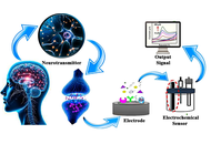
Advancements in electrochemical sensors for neurotransmitter diagnosis: innovations in sensitivity, selectivity, and real-time monitoring
Mashrufa Akther ... Md. Mahmud Alam
Published: March 24, 2026 Explor BioMat-X. 2026;3:101363
Open Access
Review
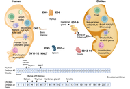
Advancing infectious disease research through a chicken embryo model for developmental studies and antimicrobial screening of lipid nanoparticles
Marta Ruano ... Valerie A. Ferro
Published: March 08, 2026 Explor BioMat-X. 2026;3:101362
Open Access
Original Article
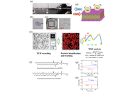
Direct observation of thermally induced fluctuation in lipid membranes using TEM-based gold nanoparticle tracking
Kazuhiro Mio ... Yuji C. Sasaki
Published: March 04, 2026 Explor BioMat-X. 2026;3:101361
Open Access
Perspective
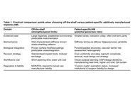
The customization paradox: Why geometric precision is no substitute for biological integration?
Frank Traub ... Felix Wunderlich
Published: March 02, 2026 Explor BioMat-X. 2026;3:101360
This article belongs to the special issue Metal 3D Printing of Biometals for Prostheses and Implants
Open Access
Perspective
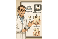
Bioprinted enamel–dentin inlays: a future direction in regenerative dentistry
Luca Fiorillo
Published: February 11, 2026 Explor BioMat-X. 2026;3:101359
This article belongs to the special issue Innovations in Biomaterials for Dentistry and Oral Surgery
Open Access
Review
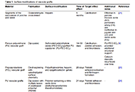
Advancements in surface modification strategies of vascular grafts to improve biocompatibility and tissue integration
Noor Abu Jarad ... Tohid F. Didar
Published: September 13, 2024 Explor BioMat-X. 2024;1:241–265
Open Access
Review
Chitosan-based composite scaffolds for accelerated epidermal-dermal wound healing
Athira S. Dev ... Renu Mohan
Published: April 24, 2025 Explor BioMat-X. 2025;2:101336
This article belongs to the special issue Nature-Based Biomaterials for Biomedical Applications
Open Access
Review
Metal 3D printing of biometals for prostheses and implants: a review
Apurba Das, Pradhyut Rajkumar
Published: May 13, 2025 Explor BioMat-X. 2025;2:101338
This article belongs to the special issue Metal 3D Printing of Biometals for Prostheses and Implants
Open Access
Review
Advancements in hydrogel technology for ocular drug delivery
Doanh Truong ... Simon D. Tran
Published: December 12, 2024 Explor BioMat-X. 2024;1:331–352
This article belongs to the special issue Bioprinted Hydrogels for Engineering Tissues and Organs
Open Access
Original Article
Long-term durability of silicone breast implants
Vladimir V. Shadrin ... Violetta A. Platunova
Published: April 09, 2025 Explor BioMat-X. 2025;2:101334
This article belongs to the special issue The Use of 2D Materials in Bio-tribology
Open Access
Commentary
Navigating regulatory challenges in molecularly tailored nanomedicine
Ajay Vikram Singh ... Paolo Zamboni
Published: April 25, 2024 Explor BioMat-X. 2024;1:124–134
Open Access
Commentary
Navigating regulatory challenges in molecularly tailored nanomedicine
Ajay Vikram Singh ... Paolo Zamboni
Published: April 25, 2024 Explor BioMat-X. 2024;1:124–134
Open Access
Review
Advancements in surface modification strategies of vascular grafts to improve biocompatibility and tissue integration
Noor Abu Jarad ... Tohid F. Didar
Published: September 13, 2024 Explor BioMat-X. 2024;1:241–265
Open Access
Original Article
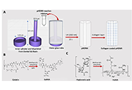
Fabrication and characterization of pHEMA hydrogel conduit containing GelMA-HaMA IPN for peripheral nerve regeneration
Damla Arslantunali Sahin ... Vasif Hasirci
Published: February 26, 2024 Explor BioMat-X. 2024;1:34–57
This article belongs to the special issue Nature-Based Biomaterials for Biomedical Applications
Open Access
Review
Chitosan-based composite scaffolds for accelerated epidermal-dermal wound healing
Athira S. Dev ... Renu Mohan
Published: April 24, 2025 Explor BioMat-X. 2025;2:101336
This article belongs to the special issue Nature-Based Biomaterials for Biomedical Applications
Open Access
Review
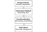
Advanced polymeric scaffolds for bone tissue regeneration
Ashkan Farazin, Seyedeh Farnaz Darghiasi
Published: June 25, 2025 Explor BioMat-X. 2025;2:101340
This article belongs to the special issue Bioinspired Material for Regenerative Medicine
Open Access
Original Article
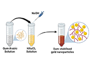
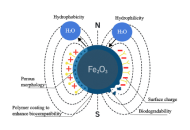
Gum Arabic-assisted green synthesis of gold nanoparticles as fluorescence modulator for potential analytical applications
Ahmed T. Algahiny ... Fryad Z. Henari
Published: July 28, 2024 Explor BioMat-X. 2024;1:190–201
This article belongs to the special issue Green Nanoparticles for Biomedical Applications
Open Access
Commentary
Navigating regulatory challenges in molecularly tailored nanomedicine
Ajay Vikram Singh ... Paolo Zamboni
Published: April 25, 2024 Explor BioMat-X. 2024;1:124–134
Open Access
Review
Advanced polymeric scaffolds for bone tissue regeneration
Ashkan Farazin, Seyedeh Farnaz Darghiasi
Published: June 25, 2025 Explor BioMat-X. 2025;2:101340
This article belongs to the special issue Bioinspired Material for Regenerative Medicine
Open Access
Review
Chitosan-based composite scaffolds for accelerated epidermal-dermal wound healing
Athira S. Dev ... Renu Mohan
Published: April 24, 2025 Explor BioMat-X. 2025;2:101336
This article belongs to the special issue Nature-Based Biomaterials for Biomedical Applications
Open Access
Review
Advancements in hydrogel technology for ocular drug delivery
Doanh Truong ... Simon D. Tran
Published: December 12, 2024 Explor BioMat-X. 2024;1:331–352
This article belongs to the special issue Bioprinted Hydrogels for Engineering Tissues and Organs
Open Access
Review
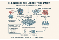
Engineering the microenvironment: advanced biomaterials for humanized in vitro immunotoxicology and carcinogenicity assessment
Sunil Choudhary ... Donato Gemmati
Published: December 08, 2025 Explor BioMat-X. 2025;2:101351
This article belongs to the special issue Bioinspired Material for Regenerative Medicine
Open Access
Review
Polymer-based nanoparticles for cancer theranostics: advances, challenges, and future perspectives
Kaylin Shanahan ... Wanis Nafo
Published: July 30, 2025 Explor BioMat-X. 2025;2:101342
Special Issues
Ongoing Special lssues
Completed Special lssues
Bioprinted Hydrogels for Engineering Tissues and Organs
Guest Editors: Vahid Serpooshan; Liqun Ning
Submission Deadline: June 30, 2026
Published Articles: 1
Green Nanoparticles for Biomedical Applications
Guest Editor: Priyanka Singh
Submission Deadline: June 30, 2026
Published Articles: 3
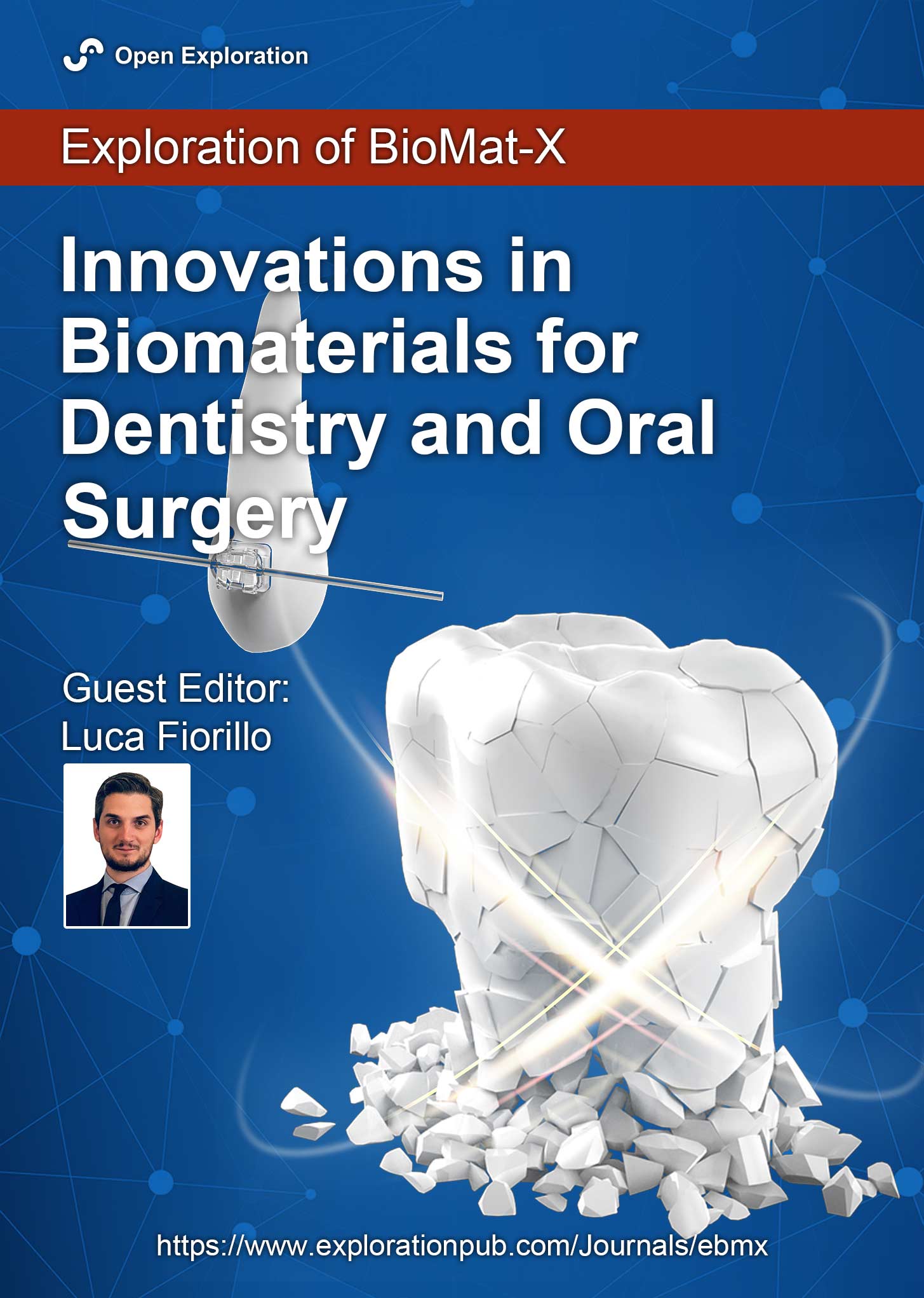
Innovations in Biomaterials for Dentistry and Oral Surgery
Guest Editor: Luca Fiorillo
Submission Deadline: June 30, 2026
Published Articles: 3
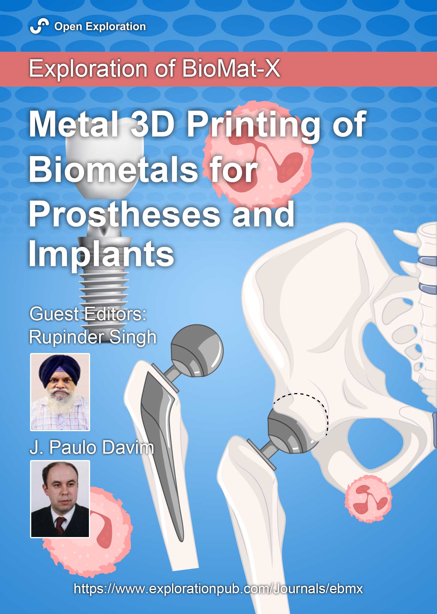
Metal 3D Printing of Biometals for Prostheses and Implants
Guest Editors: Rupinder Singh; J. Paulo Davim
Submission Deadline: June 30, 2026
Published Articles: 7
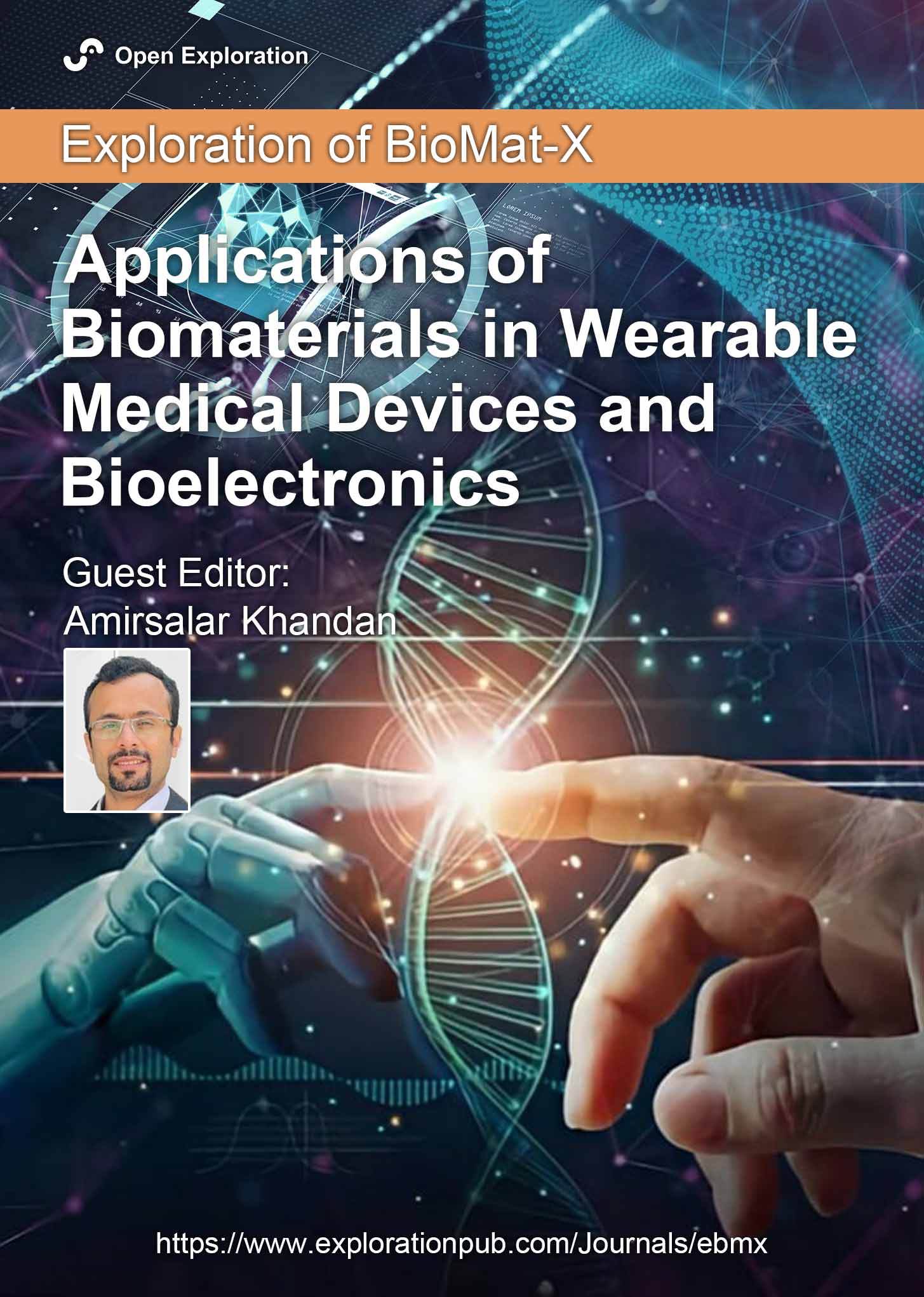
Applications of Biomaterials in Wearable Medical Devices and Bioelectronics
Guest Editor: Amirsalar Khandan
Submission Deadline: April 30, 2026
Published Articles: 0
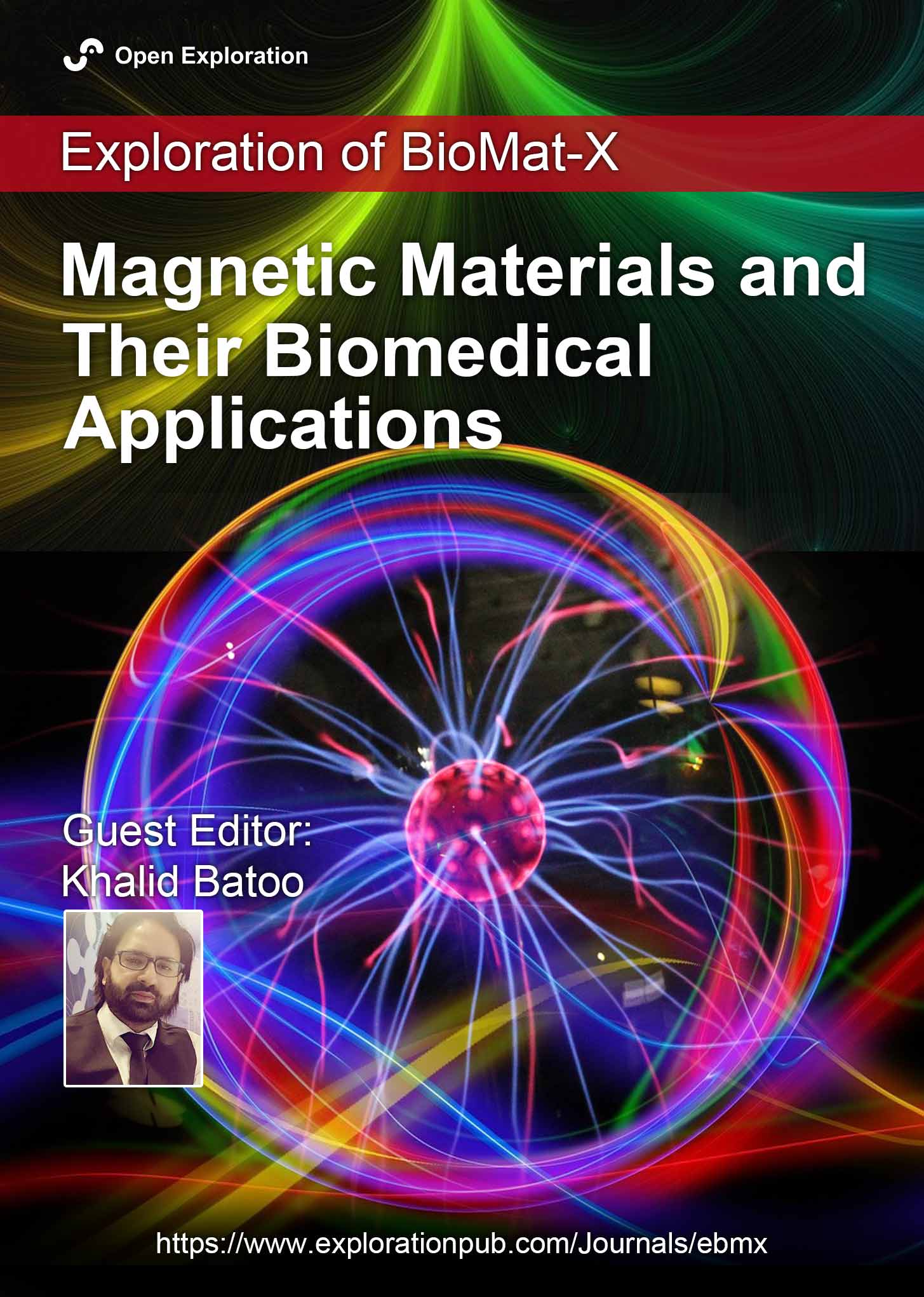
Magnetic Materials and Their Biomedical Applications
Guest Editor: Khalid Batoo
Submission Deadline: April 30, 2026
Published Articles: 0
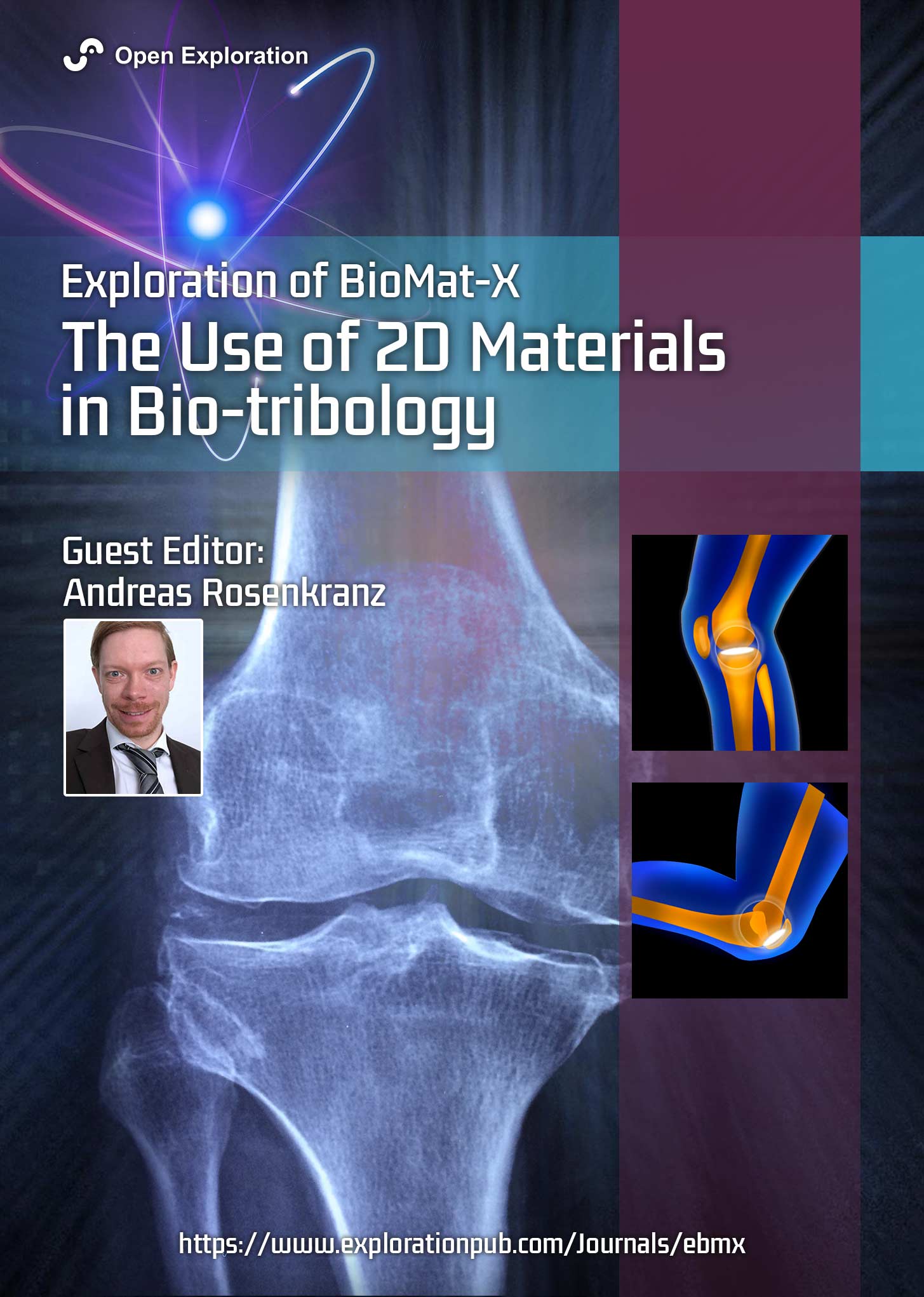
The Use of 2D Materials in Bio-tribology
Guest Editor: Andreas Rosenkranz
Submission Deadline: April 30, 2026
Published Articles: 1
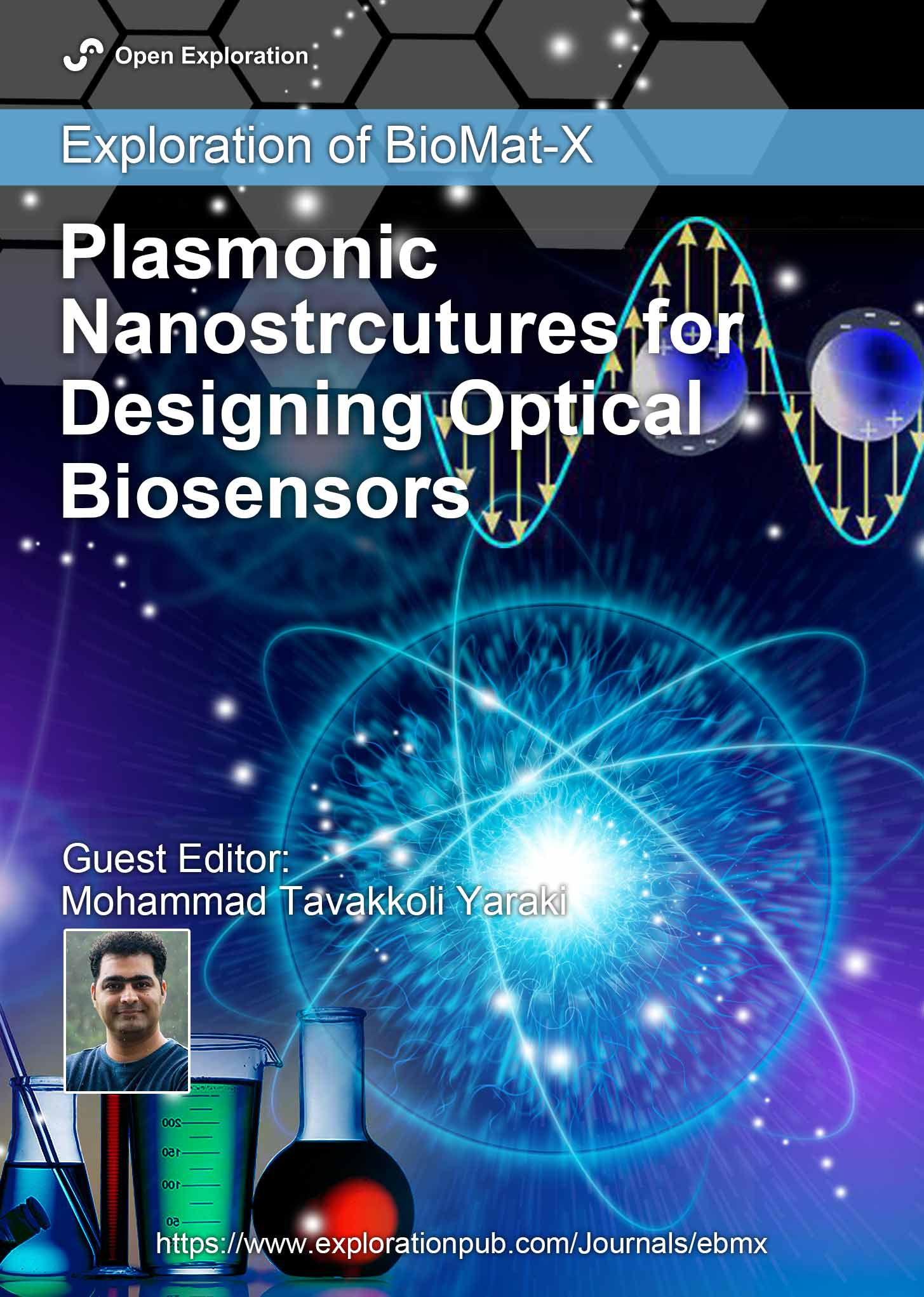
Plasmonic Nanostructures for Designing Optical Biosensors
Guest Editor: Mohammad Tavakkoli Yaraki
Submission Deadline: April 30, 2026
Published Articles: 2
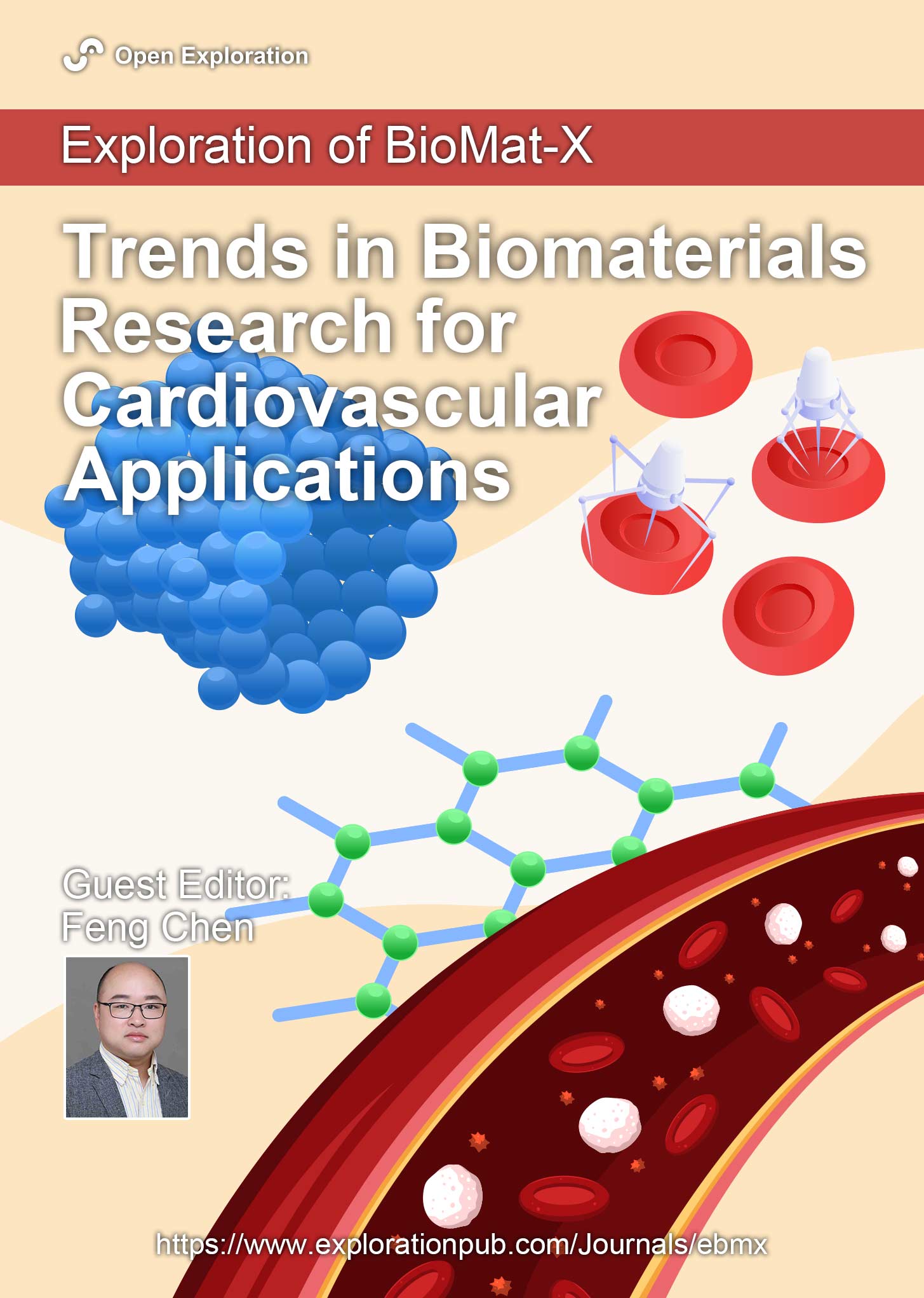
Trends in Biomaterials Research for Cardiovascular Applications
Guest Editor: Feng Chen
Submission Deadline: April 30, 2026
Published Articles: 2
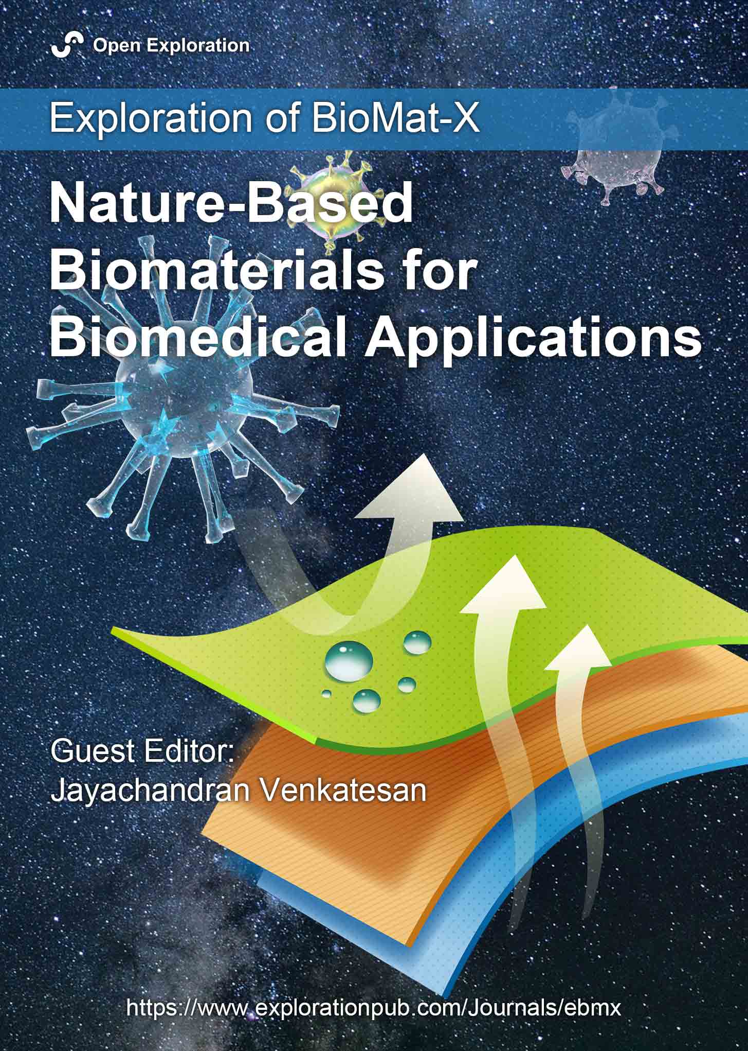
Nature-Based Biomaterials for Biomedical Applications
Guest Editor: Jayachandran Venkatesan
Submission Deadline: April 30, 2026
Published Articles: 5
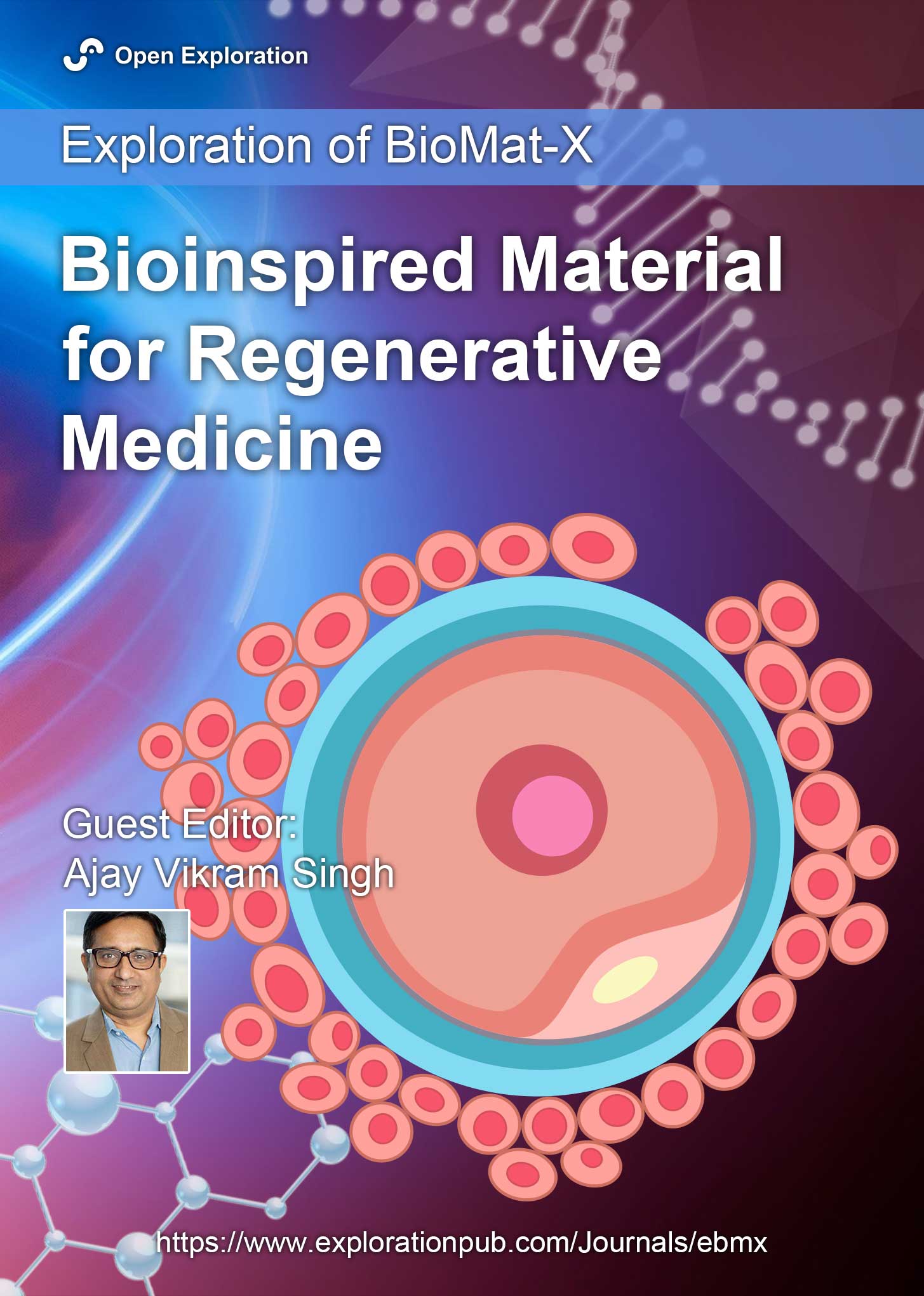
Bioinspired Material for Regenerative Medicine
Guest Editor: Ajay Vikram Singh
Submission Deadline: March 31, 2026
Published Articles: 6
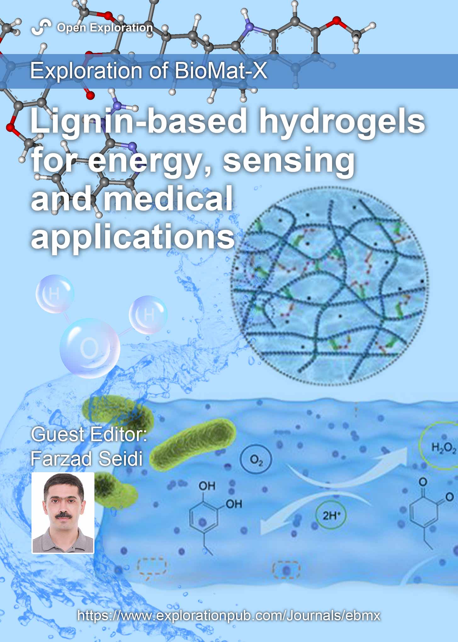
Lignin-based Hydrogels for Energy, Sensing and Medical Applications
Guest Editor: Farzad Seidi
Submission Deadline: April 30, 2026
Published Articles: 0
Membership


Exploration of Medicine is now an IUASP member journal, championing the principle that "Review work deserves to be paid". Participate in submission and peer review activities through IUASP to enjoy individual membership benefits !
Explore MembershipJournal Information
Journal Indexing
Journal Metrics
Speed


From Submission to First Decision: 5 days
From First Decision to Acceptance: 93.5 days
From Acceptance to Publication: 9 days
Article Usage (total)
Views: 354,918
Downloads: 6,547
Acceptance Rate
30.3%; 2025
56%; 2024
31.25%; 2023

 Title: Unravelling the interplaybetween #Harmattan wind andbaroreflex functions: implicationon environmental health andcardiovascular #pathophys
Follow the Journal