Open Access
Perspective
SVZonChip: a paradigm shift in hydrocephalus research and treatment
Ioannis Angelopoulos
Published: July 23, 2025 Explor Neurosci. 2025;4:1006100

Open Access
Editorial
Cutting edges in neuroscience to exceed borders
Dirk M. Hermann
Published: September 01, 2022 Explor Neurosci. 2022;1:1–3
Open Access
Review
Modify gut microbiome in autism: a promising strategy?
Jean Demarquoy ... Caroline Demarquoy
Published: August 31, 2023 Explor Neurosci. 2023;2:140–152
Open Access
Case Report
Covered stent graft for treatment of carotid artery stenosis with post-stenotic aneurysm
Mosaad Soliman ... Reem Soliman
Published: August 31, 2023 Explor Neurosci. 2023;2:153–159
Open Access
Review
Multiple sclerosis with comorbidity depression and its association with vitamin D deficiency in a narrative review of the current literature
Hans-Klaus Goischke
Published: August 31, 2023 Explor Neurosci. 2023;2:160–192
This article belongs to the special issue Novel Therapeutic Approaches for the Treatment of Depression
Open Access
Review
Negative environmental influences on the developing brain mediated by epigenetic modifications
Maya Komar-Fletcher ... Joanna Michalina Jurek
Published: September 28, 2023 Explor Neurosci. 2023;2:193–211

Open Access
Review
Sleep disorders contribute to the development of dementia and Alzheimer’s disease
Janusz Wiesław Błaszczyk
Published: October 08, 2023 Explor Neurosci. 2023;2:212–223
This article belongs to the special issue Alzheimer’s Disease
Open Access
Review
Investigation into the vascular contributors to dementia and the associated treatments
Caroline Grace Davidson ... Brandon Lucke-Wold
Published: October 15, 2023 Explor Neurosci. 2023;2:224–237
Open Access
Original Article
Estimation of the allelic status of apolipoprotein E4 isoforms with fully automated LUMIPULSE® assays
Tatsushi Yuri ... Hisashi Nojima
Published: October 16, 2023 Explor Neurosci. 2023;2:238–244
This article belongs to the special issue Alzheimer's Disease

Open Access
Mini Review
Nutritional treatment with the ketogenic diet in children with refractory epilepsy: a narrative review
Srilaxmi Vityala ... Swathi Nenavath
Published: October 30, 2023 Explor Neurosci. 2023;2:245–250
This article belongs to the special issue Epilepsy
Open Access
Review
Impact of circadian clock dysfunction on human health
Saptadip Samanta, Sk Asif Ali
Published: September 29, 2022 Explor Neurosci. 2022;1:4–30
This article belongs to the special issue Circadian Rhythm and Melatonin
Open Access
Review
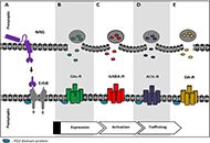
Neuregulins: subcellular localization, signaling pathways and their relationship with neuroplasticity and neurological diseases
Marines Longart ... Juan Carlos Martínez
Published: September 29, 2022 Explor Neurosci. 2022;1:31–53

Open Access
Perspective
Recent developments and future perspectives of neuropathology
Kurt A. Jellinger
Published: September 30, 2022 Explor Neurosci. 2022;1:54–60
Open Access
Review
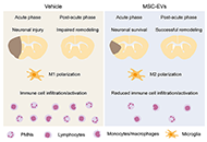
Neuroprotective and neurorestorative actions of mesenchymal stromal cell-derived small extracellular vesicles in the ischemic brain
Chen Wang ... Dirk M. Hermann
Published: October 09, 2022 Explor Neurosci. 2022;1:61–74

Open Access
Perspective
Should we rethink neurodegeneration?
Jussi O. T. Sipilä
Published: December 26, 2022 Explor Neurosci. 2022;1:75–82
Open Access
Review
Updates in mechanical thrombectomy
Kevin Pierre ... Brandon Lucke-Wold
Published: December 30, 2022 Explor Neurosci. 2022;1:83–99
Open Access
Review
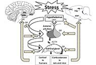
Cellular and molecular mechanisms of stress-induced memory impairment
Ameneh Rezayof ... Shiva Hashemizadeh
Published: December 30, 2022 Explor Neurosci. 2022;1:100–119
Open Access
Review
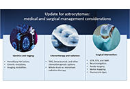
Update for astrocytomas: medical and surgical management considerations
Matthew Willman ... Brandon Lucke-Wold
Published: February 23, 2023 Explor Neurosci. 2023;2:1–26
Open Access
Systematic Review
Genetic plasma biomarkers associated with ischemic stroke
Mihai Andrei Ruscu ... Aurel Popa-Wagner
Published: February 26, 2023 Explor Neurosci. 2023;2:27–47
This article belongs to the special issue Cerebral Ischemia, Genetics, Comorbidities, Risk Factors and New Therapeutic Options for Neurorestoration

Open Access
Editorial
Treatment concept successfully translated into human patients
Dirk M. Hermann
Published: February 28, 2023 Explor Neurosci. 2023;2:48–51

