Is there a place for magnetic resonance imaging in diffuse idiopathic skeletal hyperostosis?
Diffuse idiopathic skeletal hyperostosis (DISH) is a systemic condition characterized by the new bone formation and enthesopathies of the axial and peripheral skeleton. The diagnosis of DISH current
[...] Read more.
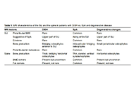
Diffuse idiopathic skeletal hyperostosis (DISH) is a systemic condition characterized by the new bone formation and enthesopathies of the axial and peripheral skeleton. The diagnosis of DISH currently relies upon the end-stage radiographic criteria of Resnick and Niwayama, in which bridging osteophytes are present over at least four thoracic vertebras. The pathogenesis of DISH is not well understood, and it is currently considered a non-inflammatory condition with an underlying metabolic derangement. However, an inflammatory component was suggested due to the similarities between DISH and spondyloarthritis (SpA) in spinal and peripheral entheseal new bone formation. Magnetic resonance imaging (MRI) is the imaging modality of choice in the diagnostic work-up and follow-up of patients with SpA, as well as in understanding its pathogenesis. The aims of the current review were to evaluate the current and future role of MRI in imaging DISH.
Iris Eshed
Diffuse idiopathic skeletal hyperostosis (DISH) is a systemic condition characterized by the new bone formation and enthesopathies of the axial and peripheral skeleton. The diagnosis of DISH currently relies upon the end-stage radiographic criteria of Resnick and Niwayama, in which bridging osteophytes are present over at least four thoracic vertebras. The pathogenesis of DISH is not well understood, and it is currently considered a non-inflammatory condition with an underlying metabolic derangement. However, an inflammatory component was suggested due to the similarities between DISH and spondyloarthritis (SpA) in spinal and peripheral entheseal new bone formation. Magnetic resonance imaging (MRI) is the imaging modality of choice in the diagnostic work-up and follow-up of patients with SpA, as well as in understanding its pathogenesis. The aims of the current review were to evaluate the current and future role of MRI in imaging DISH.