Methods:
This was a case-control study using data from the Genetics of Osteoarthritis and Lifestyle (GOAL) study. Radiographic measurements at the knee were undertaken by a single trained observer. Measurement of 12 characteristics was undertaken in 815 controls with asymptomatic structurally normal knees to examine right-left symmetry and variation with gender and age. Measurements were then compared to “cases” (315 asymptomatic and structurally unaffected knees of people with radiographic and symptomatic OA in the contralateral knee) on the assumption that the morphology of the unaffected knee represented the morphology of the contralateral knee prior to the development of OA. Right-left symmetry of morphological measures in controls was examined using paired t test and minimal detectable change (MDC). Linear regression was used to examine the association between measurements and demographic characteristics. Association of morphological features and unilateral KOA [defined as OA in either patellofemoral (PF) or tibiofemoral (TF) joints], PFOA and TFOA were determined using binary logistic regression and odds ratio (OR) and 95% confidence interval (CI) calculated. Cumulative risk of measurements in determining OA was examined using receiver operating characteristic (ROC) curves.
Results:
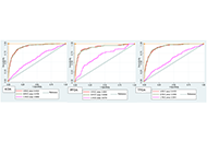
Narrow sulcus and condylar angles, increasing distal femoral, proximal tibial tilt, and increasing varus alignment associated with KOA. ROC curves including all significant morphological features and age, gender, height, and weight predicted knee, PF joint (PFJ), and TF joint (TFJ) OA with area under the curve (AUC) of 0.91, 0.89, and 0.90 respectively. On the contrary, a model only containing age, gender, height, and weight predicted knee, PFJ, and TFJ OA with AUC of 0.59, 0.67, and 0.59 respectively.
 Morphological variations at the knee associated with osteoarthritis: a case-control study using data from the GOAL studyOpen AccessOriginal ArticleAim: To identify constitutional morphological features at the knee that associate with knee osteoarthritis (OA, KOA). Methods: This was a case-control study using data from the Genetics of [...] Read more.Anand Ramachandran Nair ... Abhishek AbhishekPublished: June 30, 2023 Explor Musculoskeletal Dis. 2023;1:68–76
 Patient self-sampling for remote human leucocyte antigen-B27 analysisOpen AccessLetter to the EditorHannah Labinsky ... Johannes KnitzaPublished: June 30, 2023 Explor Musculoskeletal Dis. 2023;1:64–67
 Quantitative magnetic resonance spectroscopy and imaging analysis of the lipid content in the psoas major and its association with intervertebral disc degeneration: a cross-sectional studyOpen AccessOriginal ArticleAim: It is shown that the diminished function of the psoas major is mainly associated with increased lipid content; nonetheless, whether the fat content of the psoas major is associated with inte [...] Read more.Izaya Ogon ... Atsushi TeramotoPublished: June 29, 2023 Explor Musculoskeletal Dis. 2023;1:54–63