Non-alcoholic fatty liver disease and transient elastography
Nonalcoholic fatty liver disease (NAFLD) is a serious condition that can lead to fibrosis, cirrhosis, and hepatocellular carcinoma. NAFLD is associated with metabolic syndrome (MetS) and all of its components. According to data, a
[...] Read more.
Nonalcoholic fatty liver disease (NAFLD) is a serious condition that can lead to fibrosis, cirrhosis, and hepatocellular carcinoma. NAFLD is associated with metabolic syndrome (MetS) and all of its components. According to data, around 25–30% of population has NAFLD. Giving the growing incidence of MetS, obesity and diabetes mellitus type 2, NAFLD related terminal-stage liver disease is becoming prevailing indication for liver transplantation. In order to prevent terminal stage of this disease, it is crucial to determine those that are in risk group, to modify their risk factors and monitor their potential progression. In the absence of other causes of chronic liver disease, the prime diagnosis of NAFLD in daily clinical practice includes anamnesis, laboratory results (increased levels of aminotransferases and gammaglutamil transferases) and imaging methods. The biggest challenge with NAFLD patients is to differentiate simple steatosis from nonalcoholic steatohepatitis, and detection of fibrosis, that is the main driver in NAFLD progression. The gold standard for NAFLD diagnosis still remains the liver biopsy (LB). However, in recent years many noninvasive methods were invented, such as transient elastography (TE). TE (FibroScan®, Echosens, Paris, France) is used for diagnosis of pathological differences of liver stiffness measurement (LSM) and controlled attenuation parameter (CAP). Investigations in the last years have confirmed that elastographic parameters of steatsis (CAP) and fibrosis (LSM) are reliable biomarkers to non-invasively assess liver steatosis and fibrosis respectively in NAFLD patients. A quick, straightforward and non-invasive method for NAFLD screening in patients with MetS components is TE-CAP. Once diagnosed, the next step is to determine the presence of fibrosis by LSM which should point out high risk patients. Those patients should be referred to hepatologists. LB may be avoided in a substantial number of patients if TE with CAP is used for screening.
Ivana Mikolasevic ... Sandra Milic
Nonalcoholic fatty liver disease (NAFLD) is a serious condition that can lead to fibrosis, cirrhosis, and hepatocellular carcinoma. NAFLD is associated with metabolic syndrome (MetS) and all of its components. According to data, around 25–30% of population has NAFLD. Giving the growing incidence of MetS, obesity and diabetes mellitus type 2, NAFLD related terminal-stage liver disease is becoming prevailing indication for liver transplantation. In order to prevent terminal stage of this disease, it is crucial to determine those that are in risk group, to modify their risk factors and monitor their potential progression. In the absence of other causes of chronic liver disease, the prime diagnosis of NAFLD in daily clinical practice includes anamnesis, laboratory results (increased levels of aminotransferases and gammaglutamil transferases) and imaging methods. The biggest challenge with NAFLD patients is to differentiate simple steatosis from nonalcoholic steatohepatitis, and detection of fibrosis, that is the main driver in NAFLD progression. The gold standard for NAFLD diagnosis still remains the liver biopsy (LB). However, in recent years many noninvasive methods were invented, such as transient elastography (TE). TE (FibroScan®, Echosens, Paris, France) is used for diagnosis of pathological differences of liver stiffness measurement (LSM) and controlled attenuation parameter (CAP). Investigations in the last years have confirmed that elastographic parameters of steatsis (CAP) and fibrosis (LSM) are reliable biomarkers to non-invasively assess liver steatosis and fibrosis respectively in NAFLD patients. A quick, straightforward and non-invasive method for NAFLD screening in patients with MetS components is TE-CAP. Once diagnosed, the next step is to determine the presence of fibrosis by LSM which should point out high risk patients. Those patients should be referred to hepatologists. LB may be avoided in a substantial number of patients if TE with CAP is used for screening.
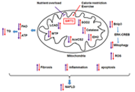 Role of acetylation in nonalcoholic fatty liver disease: a focus on SIRT1 and SIRT3Open AccessReviewNonalcoholic fatty liver disease (NAFLD) has become the most prevalent liver chronic disease worldwide. The pathogenesis of NAFLD is complex and involves many metabolic enzymes and multiple pathways. Posttranslational modification [...] Read more.Fatiha NassirPublished: August 31, 2020 Explor Med. 2020;1:248–258
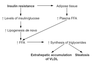 Non-alcoholic fatty liver disease and transient elastographyOpen AccessReviewNonalcoholic fatty liver disease (NAFLD) is a serious condition that can lead to fibrosis, cirrhosis, and hepatocellular carcinoma. NAFLD is associated with metabolic syndrome (MetS) and all of its components. According to data, a [...] Read more.Ivana Mikolasevic ... Sandra MilicPublished: August 31, 2020 Explor Med. 2020;1:205–217
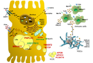 Genetic and metabolic factors: the perfect combination to treat metabolic associated fatty liver diseaseOpen AccessReviewThe prevalence of nonalcoholic or more recently re-defined metabolic associated fatty liver disease (MAFLD) is rapidly growing worldwide. It is characterized by hepatic fat accumulation exceeding 5% of liver weight not attributabl [...] Read more.Marica Meroni ... Paola DongiovanniPublished: August 31, 2020 Explor Med. 2020;1:218–243
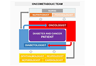 Type 2 diabetes and cancer: problems and suggestions for best patient managementOpen AccessReviewDiabetes and cancer are widespread worldwide and the number of subjects presenting both diseases increased over the years. The management of cancer patients having diabetes represents a challenge not only because of the complexity [...] Read more.Agostino Milluzzo ... Laura SciaccaPublished: August 31, 2020 Explor Med. 2020;1:184–204
 Visual communication and learning from COVID-19 to advance preparedness for pandemicsOpen AccessPerspectiveThe currently ongoing coronavirus disease 19 (COVID-19) pandemic has affected globally human health and economy. Research in progress has shown facts associated with this disease and raised questions relevant for disease control a [...] Read more.José de la Fuente ... Christian GortázarPublished: August 31, 2020 Explor Med. 2020;1:244–247
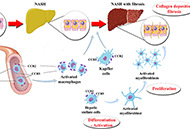 The therapeutic potential of C-C chemokine receptor antagonists in nonalcoholic steatohepatitisOpen AccessReviewPooled prevalence of nonalcoholic fatty liver disease (NAFLD) globally is about 25%. Nonalcoholic steatohepatitis (NASH) with advanced fibrosis has been linked with substantial morbidity and mortality, without having to-date any l [...] Read more.Michael Doulberis ... Stergios A. PolyzosPublished: August 31, 2020 Explor Med. 2020;1:170–183